

Abstract
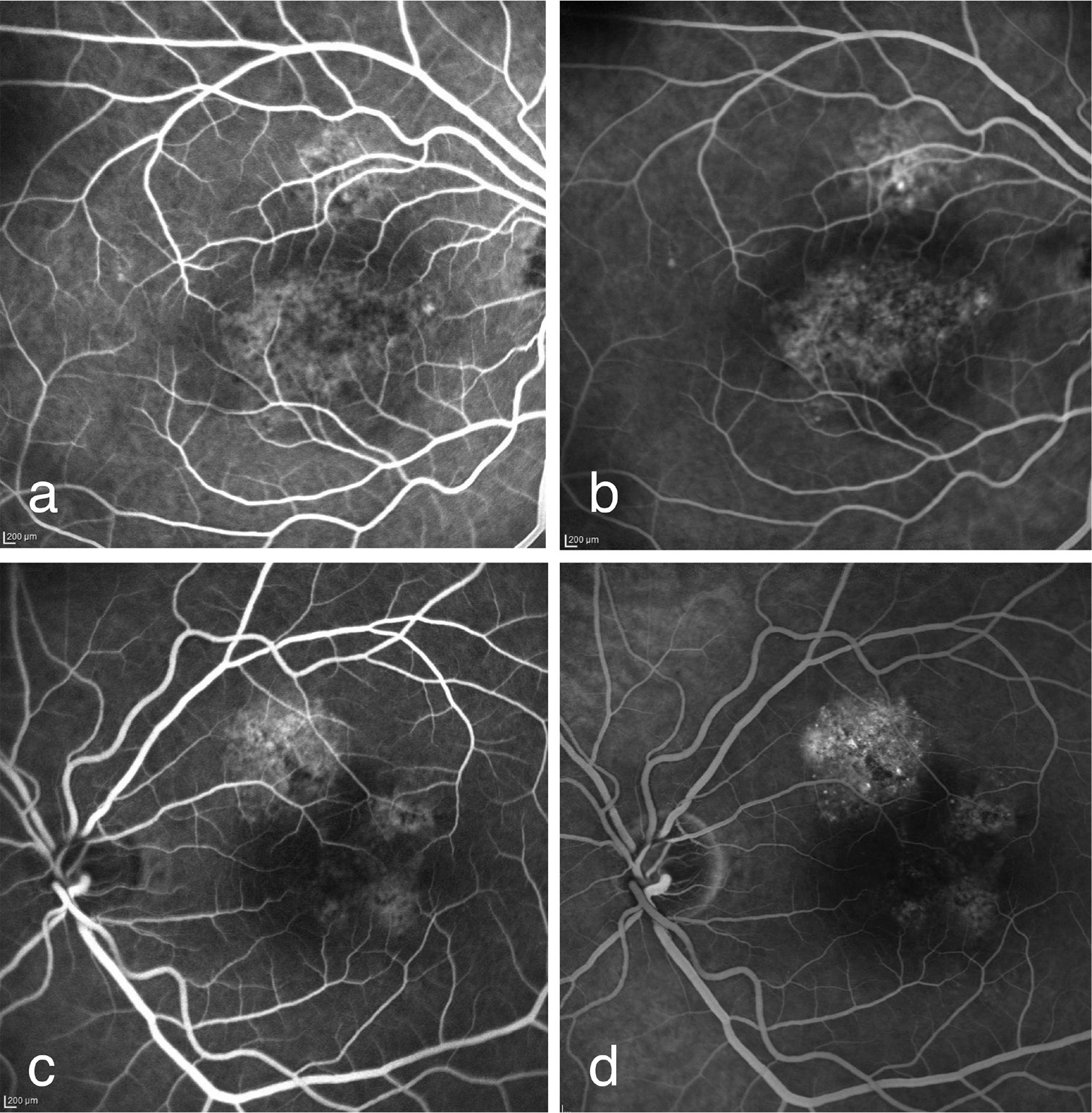
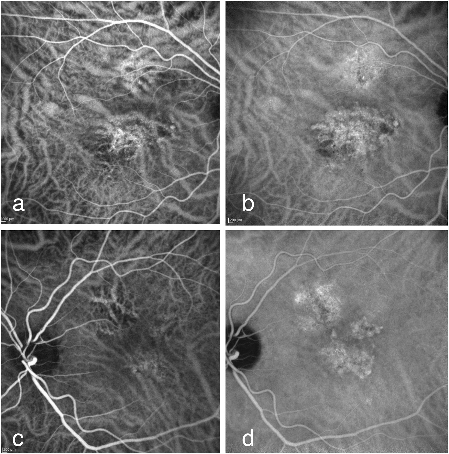
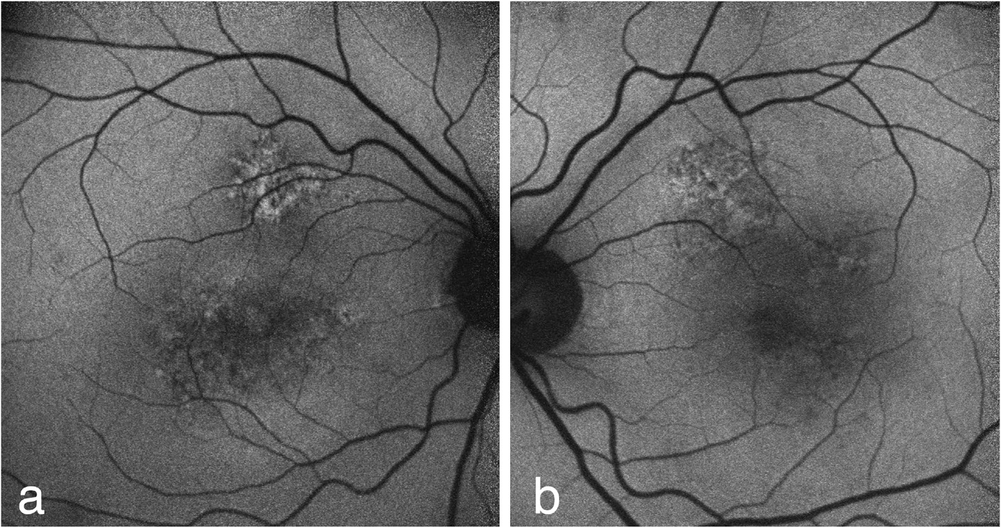
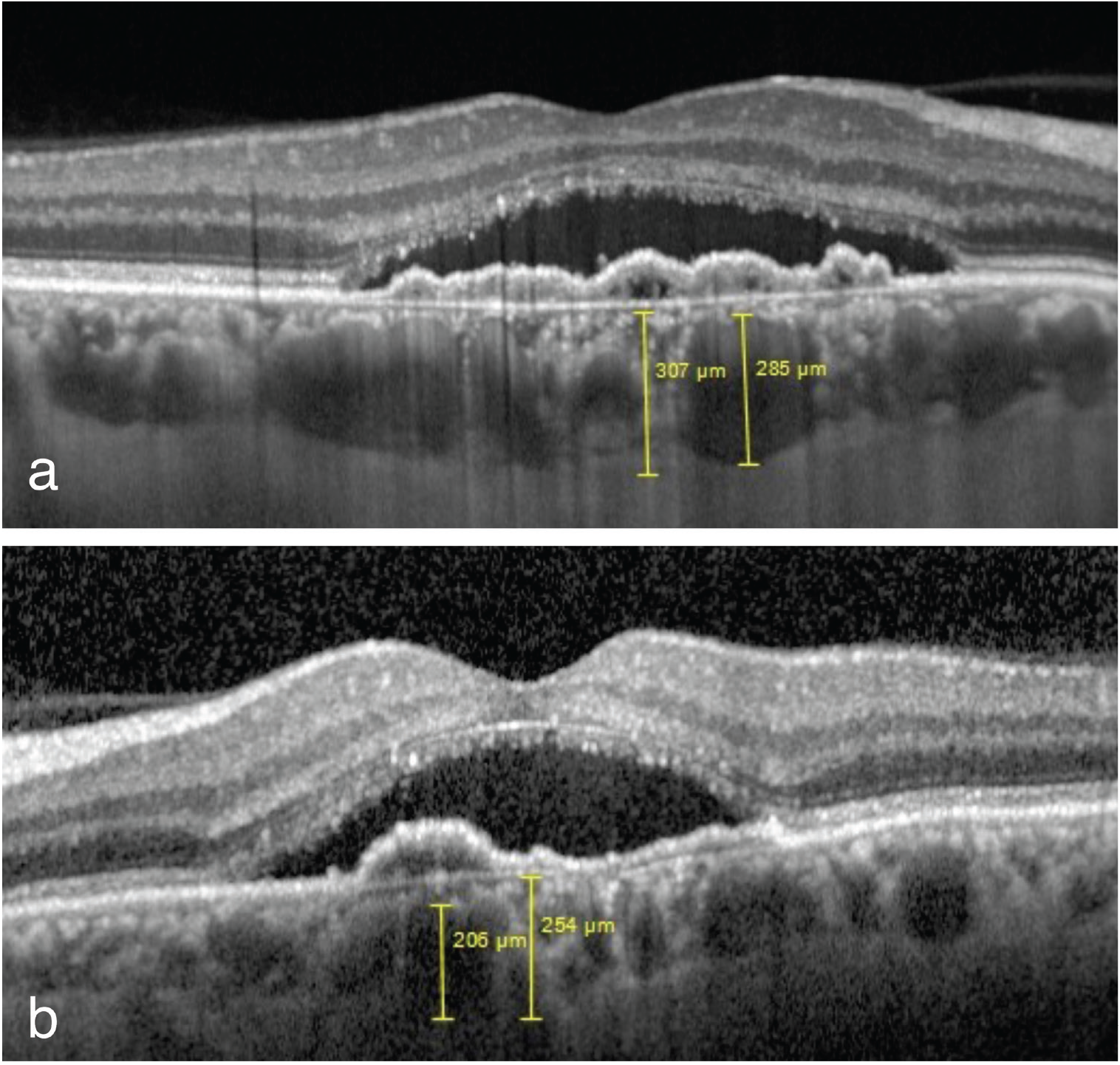
Pachychoroid neovasculopathy (PNV) is a form of type 1 neovascularization characterized by dilated choroidal vessels in areas of increased choroidal thickness. In this article, we describe a patient diagnosed with PNV. A 50-year-old male with a 2-month history of blurred vision was referred to our clinic. His best corrected visual acuity was 20/100 in both eyes. Retinal pigment epithelium alterations, which were more prominent in fundus autofluorescence, were detected in both eyes on dilated fundus examination. Characteristic findings of PNV were detected in fundus fluorescein angiography, indocyanine green angiography, spectral domain optical coherence tomography, and optical coherence tomography angiography.
Keywords: Pachychoroid neovasculopathy, choroidal neovascularization, optical coherence tomography angiography
Introduction
Pachychoroid spectrum diseases were first recognized in 2013 when Warrow et al. described pachychoroid pigment epitheliopathy. The pachychoroid spectrum includes 4 disease groups: pachychoroid pigment epitheliopathy, central serous chorioretinopathy, pachychoroid neovasculopathy (PNV), and polypoidal choroidal vasculopathy. Pachychoroid spectrum diseases are characterized by increased choroidal thickness, dilation of the outer choroidal veins (pachy-veins), and thinning of Sattler’s and choriocapillaris layers.
Multimodal imaging methods are used to understand the disease pathophysiology and in diagnosis. Indocyanine green angiography (ICGA) is shown to be superior to fundus fluorescein angiography (FFA) for detailed imaging of choroid neovascularization (CNV) and diagnosis of choroidal polyps. Thanks to its longer wavelength, ICGA enables better visualization of lesions underlying the retinal pigment epithelium (RPE), even in the presence of blood, exudate, and pigment epithelium detachment (PED)., Advances in the field of optical coherence tomography (OCT) have also enabled imaging of choroidal structures in addition to the retina., OCT angiography (OCT-A), a relatively new technology, provides structural information about the retinal and choroidal vessels without the need for contrast material injection.
In this case report, we analyze the findings obtained with various imaging modalities from a patient with PNV who presented with a 2-month history of blurred vision.
Discussion
PNV was first described by Pang and Freund in 2015. The disease may consist of type 1 CNV that develops secondary to central serous chorioretinopathy or pachychoroid pigment epitheliopathy. PNV should be suspected in cases of thickened choroid with type 1 CNV without characteristic findings of age-related macular degeneration (AMD) such as drusen or hemorrhages. It is characterized by the presence of shallow, irregular PED.
In a study by Miyake et al. including 200 patients diagnosed with PNV and AMD, 19.5% of the cases were diagnosed with PNV. Subfoveal choroid thickness was found to be greater in patients with PNV than in those with AMD. They reported that genetic mutations were detected less frequently in patients with PNV. In addition, PNV was observed in younger patients compared to AMD.
The etiopathogenesis of pachychoroid spectrum diseases involves microtrauma to the Bruch’s membrane from the enlarged pachy-veins in the Haller’s layer. This causes choriocapillaris loss and RPE changes. Neovasculopathy develops as a result of extension of neovascularization beneath the RPE.
FFA and ICGA are used in the diagnosis of CNV and are known to cause nausea and anaphylaxis in rare cases. OCT-A enables image acquisition by serial OCT scanning and is a reliable method that allows imaging of retinal and choroidal vasculature without needing any dye injection. The tangled vascular network under the shallow irregular PED can be imaged with OCT-A. In a study including 16 patients (22 eyes) with shallow irregular PED, CNV was detected in 95% of the patients with OCT-A. Compared to other angiography techniques, OCT-A is shown to be more successful in demonstrating type 1 CNV.
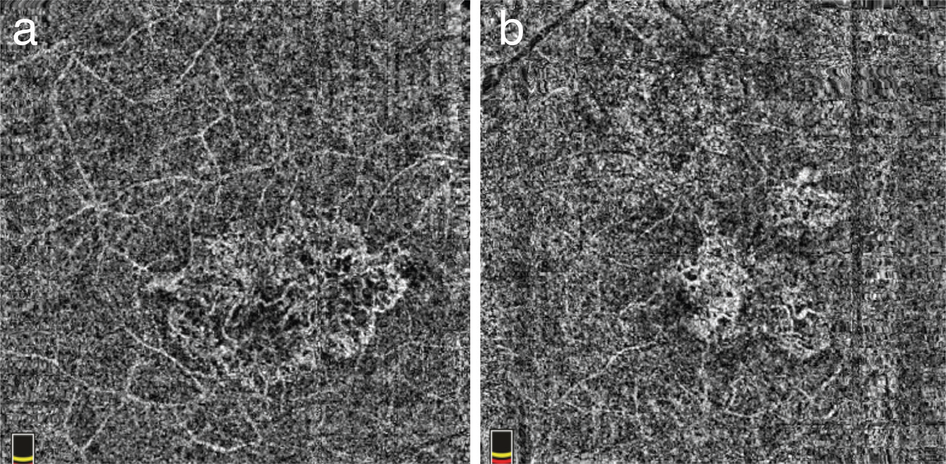
Similarly, in the present case we observed shallow PED, subretinal fluid, thickened choroid, and pachy-veins on spectral domain OCT and appearance consistent with type 1 CNV in FFA and ICGA. OCTA showed a CNV network in the areas corresponding to the type 1 CNV observed on ICGA. In a case series by Azar et al. including 5 PNV patients, the presence of neovascularization could not be fully identified with FFA and ICGA in 2 patients, whereas the presence of tangled filamentous vascular network was detected in all of the patients with OCT-A. Therefore, these findings indicate that OCT-A can detect CNV before FFA and ICGA in pachychoroid spectrum diseases.
In conclusion, as we have also observed in our case, non-invasive OCT-A imaging generally supports fundus angiography images with regard to the diagnosis of type 1 CNV in PNV. OCT-A should be used in combination with other methods for the detection of vascularization in AMD presenting with shallow PED and in pachychoroid spectrum diseases.
References
Warrow DJ, Hoang QV, Freund KB. Pachychoroid pigment epitheliopathy. Retina. 2013;33:1659–1672. [PubMed] [Google Scholar]
Dansingani KK, Balaratnasingam C, Naysan J, Freund KB. En face imaging of pachychoroid spectrum disorders with swept-source optical coherence tomography. Retina. 2016;36:499–516. [PubMed] [Google Scholar]
Spaide RF, Yanuzzi LA, Slakter JS, Sorenson J, Orlach DA. Indocyanine green videoangiography of idiopathic polypoidal choroidal vasculopathy. Retina. 1995;15:100–110. [PubMed] [Google Scholar]
Yannuzzi LA. Indocyanine green angiography: a perspective on use in the clinical setting. Am J Ophtalmol. 2011;151:745–751. [PubMed] [Google Scholar]
Spaide RF, Koizumi H, Pozzoni MC. Enhanced depth imaging spectral-domain optical coherence tomography. Am J Ophthalmol. 2008;146:496–500. [PubMed] [Google Scholar]
Potsaid B, Baumann B, Huang D, Barry S, Cable AE, Schuman JS, Duker JS, Fujimoto JG. Ultrahigh speed 1050 nm swept source/Fourier domain OCT retinal and anterior segment imaging at 100,000 to 400,000 axial scans per second. Opt Express. 2010;18:20029–20048. [PMC free article] [PubMed] [Google Scholar]
Ferrara D, Waheed NK, Duker JS. Investigating the choriocapillaris and choroidal vasculature with new optical coherence tomography technologies. Prog Retin Eye Res. 2016;52:130–155. [PubMed] [Google Scholar]
Pang CE, Freund KB. Pachychoroid neovasculopathy. Retina. 2015;35:1–9. [PubMed] [Google Scholar]
Miyake M, Ooto S, Yamashiro K, Takahashi A, Yoshikawa M, Akagi-Kurashige Y, Ueda-Arakawa N, Oishi A, Nakanishi H, Tamura H, Tsujikawa A, Yoshimura N. Pachychoroid neovasculopathy and age-related macular degeneration. Sci Rep. 2015;5:16204. [PMC free article] [PubMed] [Google Scholar]
Hata M, Yamashiro K, Ooto S, Oishi A, Tamura H, Miyata M, Ueda-Arakawa N, Takahashi A, Tsujikawa A, Yoshimura N. Intraocular Vascular Endothelial Growth Factor Levels in Pachychoroid Neovasculopathy and Neovascular Age-Related Macular Degeneration. Invest Ophthalmol Vis Sci. 2017;58:292–298. [PubMed] [Google Scholar]
Su Z, Teng Y, Zhang L, Shu X. Adverse reaction in patients with drug allergy history after simultaneous intravenous fluorescein angiography and indocyanine green angiography. J Ocul Phrmacol Ther. 2012;28:410–413. [PubMed] [Google Scholar]
Dansingani KK, Balaratnasingam C, Klufas MA, Sarraf D, Freund KB. Optical Coherence Tomography Angiography of Shallow Irregular Pigment Epithelial Detachments In Pachychoroid Spectrum Disease. Am J Ophthalmol. 2015;160:1243–1254. [PubMed] [Google Scholar]
Azar G, Wolff B, Mauget-Faÿsse M, Rispoli M, Savastano MC, Lumbroso B. Pachychoroid neovasculopathy: aspect on optical coherence tomography angiography. Acta Ophthalmol. 2017;95:421–427. [PubMed] [Google Scholar]

