

Abstract
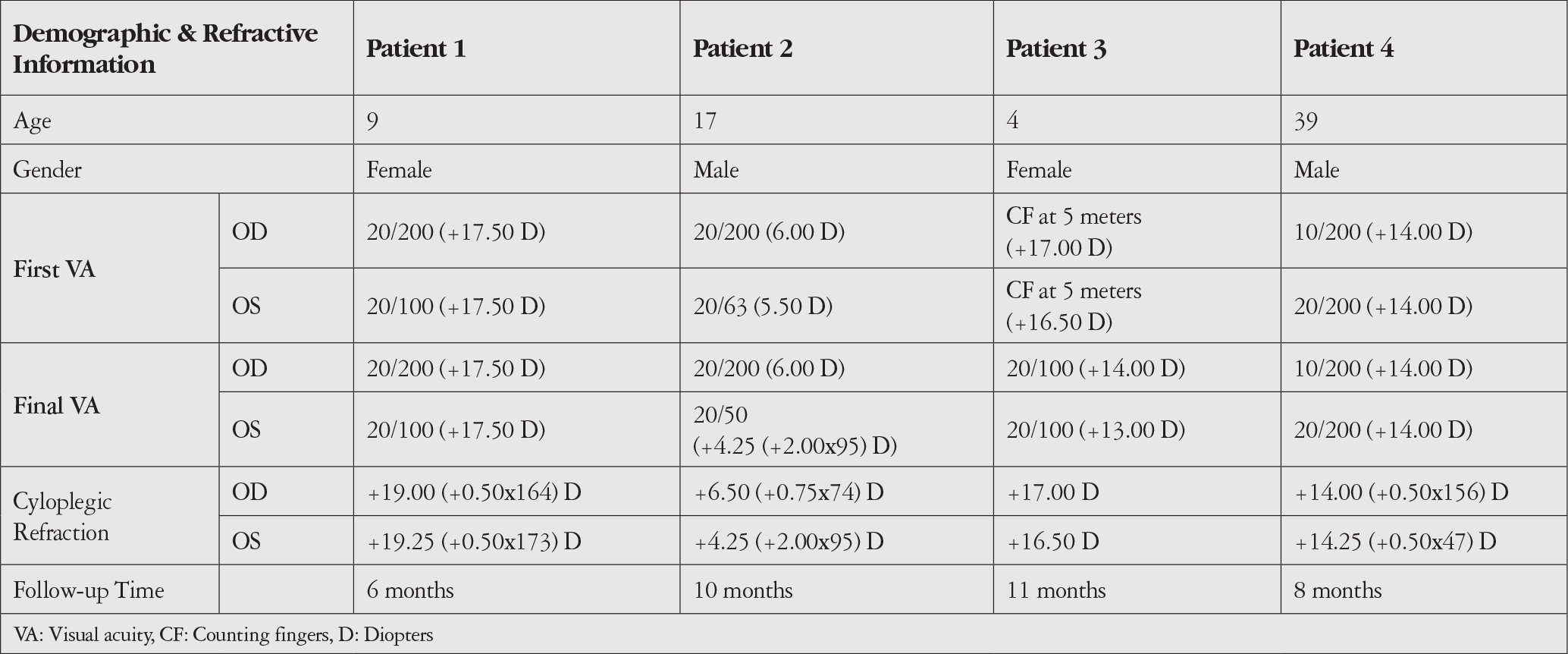
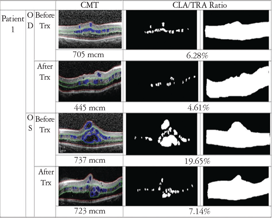
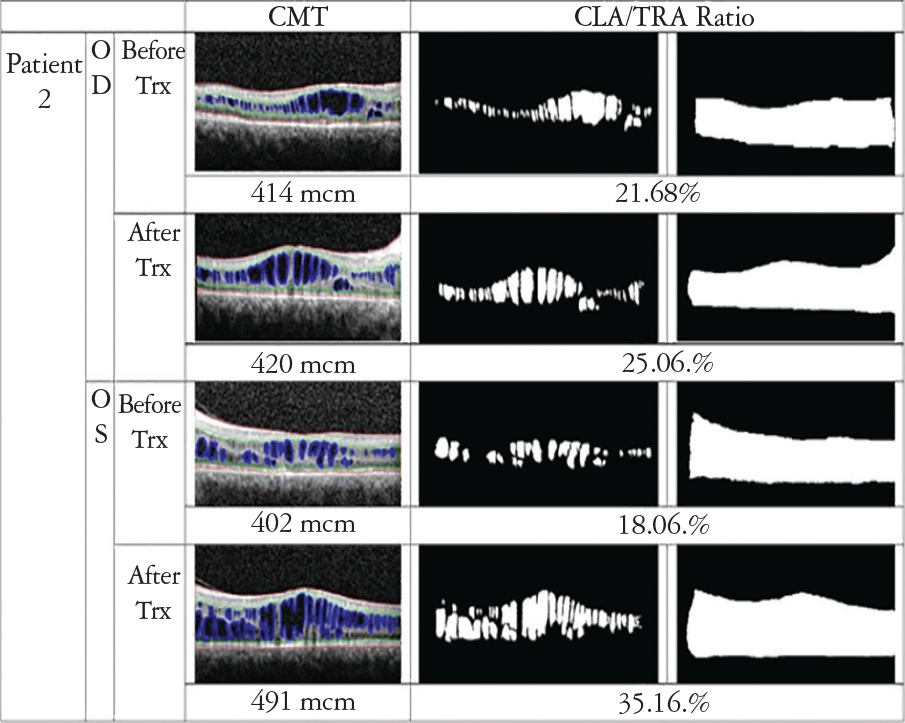
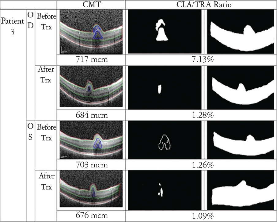
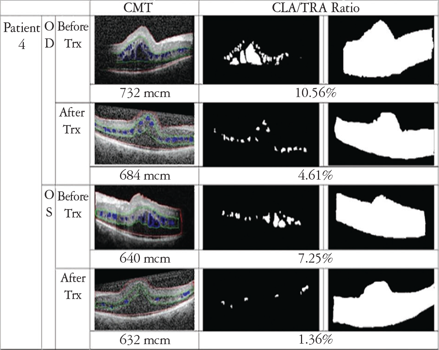
The aim of this study was to report the outcome of topical brinzolamide 1% treatment on macular cystoid lesions resembling retinoschisis in 4 patients diagnosed with posterior microphthalmia. The medical records of 4 patients with a clinical diagnosis of posterior microphthalmia who had started topical brinzolamide 1% treatment were reviewed. Visual acuity, central foveal thickness, and cystoid lesion area percentage were used to evaluate treatment response. In the follow-up, there was a decrease in central foveal thicknesses and cystoid lesion area percentages in both eyes of 3 of the patients. However, 1 patient showed increases in both parameters. Visual acuity remained stable in 5 eyes and increased in 3 eyes. Topical brinzolamide treatment may have some positive effects on macular cystoid lesions in selected cases.
Keywords: Brinzolamide, macular cystoid lesion, retinoschisis, posterior microphthalmos
Introduction
Microphthalmos is an ocular developmental disorder characterized by an eye with a total axial length of more than two standard deviations smaller than normal for that age group. Posterior microphthalmos (PM) is a type of microphthalmos which is defined as hyperopia, a short axial length, posterior segment foreshortening and a normal-appearing anterior segment of normal or subnormal dimensions.,
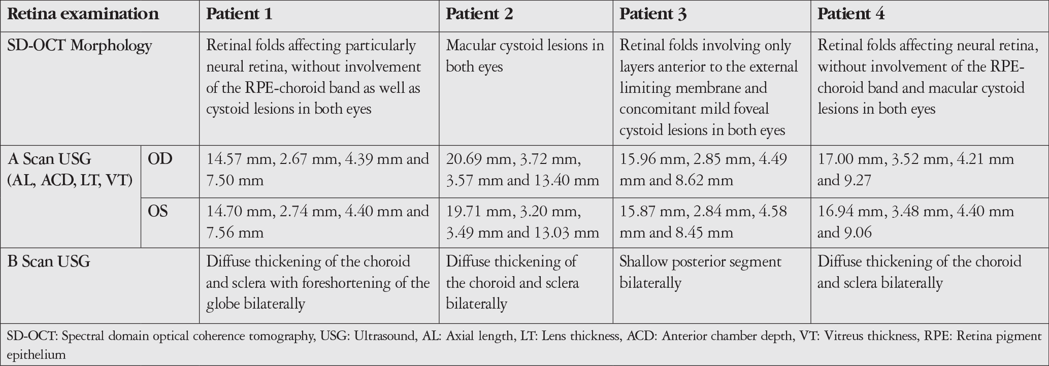
Retinal folds, macular schisis, cystoid lesions, reduced or absent foveal avascular zone, pseudopapilledema and uveal effusion are prominent posterior segment changes in PM.,,,, Optical coherence tomography (OCT) scans of PM patients demonstrate that the neurosensory retina is folded, while the retinal pigment epithelium (RPE) layer and choroid are intact without folds., The growth of neurosensory retina occurs independently of other ocular tissues, while choroid and RPE development are regulated by thickened sclera; therefore, disproportionate growth between the neurosensory retina and surrounding outer tissues causes retinal folding., Macular schisis is thought to be caused by thickened sclera, which causes blockage of the trans-scleral outflow.,
Carbonic anhydrase inhibitors (CAIs) function by acidifying the subretinal space and increasing fluid transport across the RPE; therefore, CAIs have been used for the treatment of macular schisis in various causes. In this paper, we present 4 cases of PM and evaluate the efficacy of topical brinzolamide 1% treatment on the cystoid cavities resembling retinoschisis.
Discussion
The carbonic anhydrase (CA) is a group of enzymes found extensively throughout the body. Although CA isoenzymes are diverse, the body’s most common subtype is the intracellular CA isoenzyme II. In the retina, CA II is found in the cytoplasm of red/green cones and inside the Müller cells especially. However, the RPE appears to contain the membrane-bound isoenzyme IV, which regulates the extracellular pH gradient created via the metabolic activity of cells and also acts as a bicarbonate channel.
CAIs have been proven to increase retinal adhesiveness, enhance the activity of fluid transporter on the RPE cell membrane and change subretinal acidity. The use of CAIs for retinal diseases is not uncommon. Initial investigations were conducted by Cox et al. in which 41 patients with cystoid macular edema (CME) of various causes were given oral acetazolamide. The drug was effective in more than half of the patients with uveitis or hereditary outer retinal diseases, but in none with retinal vascular disorders. According to the authors, acetazolamide increased the rate of fluorescein disappearance and fluid transport from vitreous by acting via CA-IV receptors on RPE cells. A topical CAI for a retinal disease was first used in chronic CME of retinitis pigmentosa (RP) patients and provided partial improvement in visual function. In 2006, Apushkin et al. were first to investigate the effect of a topical CAI, in particular dorzolamide 2%, on schisis and cystic macular cavities in X-linked retinoschisis (XLRS). The authors reported more than 7 letters gain in VA within 2 months in nearly half of cases.
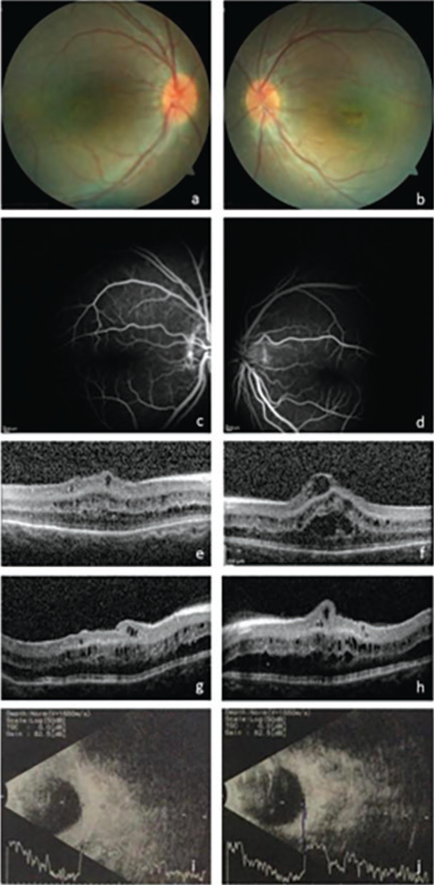
Foveoschisis is one of the most common causes of pseudo-CME. Unlike CME, there is no leakage in late phases of FFA in foveoschisis. The differential diagnosis of foveoschisis includes retinal dystrophies like XLRS, Goldmann-Favre syndrome, enhanced S-cone syndrome, and RP; high myopia; microphthalmia-nanophtalmia; and age-related retinoschisis. The cause of schisis and retinal cysts in hereditary dystrophies is thought to be associated with gene mutations (e.g., in RS1, NR2E3) causing defective protein synthesis. These proteins cause abnormal RPE and PR cell formation and ineffective cellular adhesion, and therefore splitting of the fovea. In PM, however, macular schisis is thought to arise from thickened sclera that causes impermeability of the trans-scleral outflow. Consequent congestion of the retina may result in cystic degeneration by overwhelming Müller cell function, which provides physiologic and architectural support., To the best of our knowledge, our study is first to evaluate the effect of a CAI for PM.
We investigated both the anatomical and functional outcomes of topical brinzolamide treatment. We observed a decrease in CLA percentage in both eyes of 3 patients. However, in both eyes of 1 patient, this ratio was increased. We observed that CLA percentage of this patient was higher than the other patients and cystoid macular degeneration was prominent in his OCT sections. In accordance with the literature, we hypothesized that the drug might have a greater benefit on patients with more preserved retinal anatomy. In patients with healthier RPE and Müller cells, the response to the drug will be better and the cysts can shrink more rapidly.
VA remained stable in 5 eyes and increased in 3 eyes. Reduction in CLA and CFT were not correlated with change in VA except for patient 3. Previous studies in XLRS and RP reported that reduction of CFT was not correlated with improvement in VA.,
The ages of our cases ranged from 4 to 39 years, and this difference should be considered in terms of drug efficacy and safety. In their study, Khandhadia et al. examined 4 patients diagnosed with XLRS, 3 of whom were children, and attributed the fact that the only patient who showed continuous clinical improvement with a topical CAI was the adult patient due to his potential high drug compliance. Topical brinzolamide has been reported to be effective when given twice daily in congenital glaucoma cases under 6 years of age and no serious adverse effects have been reported. Yang et al. reported that topical brinzolamide used 3 times daily was effective and safe in 4 patients aged 4 to 10 years with the diagnosis of XLRS. However, there is no comparative study between adult and pediatric age group in terms of side effect profile and posology of topical brinzolamide in the literature.
Due to potential requirement for long-term treatment, the use of oral acetazolamide may be limited by potential systemic side effects and a topical drug may be preferred. We used brinzolamide due to its better side effect profile., Karatas et al. found partial improvement in VA and relative stabilization in CFT in 8 XLRS patients with brinzolamide treatment. In their XLRS case series, Yang et al. achieved a favorable outcome with topical brinzolamide in the reduction of macular cysts, with no reported adverse drug effects. None of our patients reported any unfavorable side effects.
Conclusion
Considering the efficacy of topical brinzolamide in the treatment of macular cysts and schisis in XLRS and RP, we have tried to find a cure with brinzolamide in macular cystoid lesions resembling macular schisis of PM in which no treatment is available so far. We observed that topical brinzolamide 1% treatment caused partial reduction in cystoid lesions’ area in PM patients. Prospective studies with longer follow-up period and higher number of patients are needed for evaluating the long-term efficacy.
References
Bardakjian T, Schneider A, Weiss A. Microphthalmia/Anophthalmia/Coloboma Spectrum. In: Adam MP, Ardinger HH, Pagon RA, Wallace SE, Bean LJH, Stephens K, Amemiya A, editors. GeneReviews. Seattle: University of Washington; 2007.
Spitznas M, Gerke E, Bateman VB. Hereditary posterior microphthalmos with papillomacular fold and high hyperopia. Arch Ophthalmol. 1983;101:413-417.
Khairallah M, Messaoud R, Zaouali S, Yahia SB, Ladjimi A, Jenzri S. Posterior segment changes associated with posterior microphthalmos. Ophthalmology. 2002;109:569-574.
Alkin Z, Ozkaya A, Karakucuk Y, Demirok A. Detailed ophthalmologic evaluation of posterior microphthalmos. Middle East Afr J Ophthalmol. 2014;21:186-188.
Nguyen AT, Johnson MA, Hutcheson KA. Good visual function in posterior microphthalmos. J AAPOS. 2000;4:240-242.
Tekin K, Teke MY, Citirik M. Clinical appraisal and retinal imaging in posterior microphthalos. Semin Ophthalmol. 2018;33:412-418.
Kim JW, Boes DA, Kinyoun JL. Optical coherence tomography of bilateral posterior microphthalmos with papillomacular fold and novel features of retinoschisis and dialysis. Am J Ophthalmol. 2004;138:480-481.
Aras C, Ozdamar A, Ustundag C, Ozkan S. Optical coherence tomographic features of papillomacular fold in posterior microphthalmos. Retina. 2005;25:665-667.
Warburg M. Classification of microphthalmos and coloboma. J Med Genet. 1993;30:664-669.
Wright, Kenneth W, Spiegel PH. Pediatric Ophthalmology and Strabismus. 2nd ed. New York: Springer-Verlag. 2013:12-13.
Park SH, Ahn YJ, Shin SY, Lee YC. Clinical features of posterior microphthalmos associated with papillomacular fold and high hyperopia. Clin Exp Optom. 2016;99:590-593.
Dhrami-Gavazi E, Schiff WM, Barile GR. Nanophthalmos and acquired retinoschisis. Am J Ophthalmol. 2009;147:108-110.
Wolfensberger TJ. The role of carbonic anhydrase inhibitors in the management of macular edema. Doc Ophthalmol. 1999;97:387-397.
Nowilaty SR, Khan AO, Aldahmesh MA, Tabbara KF, Al-Amri A, Alkuraya FS. Biometric and molecular characterization of clinically diagnosed posterior microphthalmos. Am J Ophthalmol. 2013;155:361-372.e7.
Wilkins GR, Houghton OM, Oldenburg AL. Automated segmentation of intraretinal cystoid fluid in optical coherence tomography. IEEE Trans Biomed Eng. 2012;59:1109-1114.
Slokom N, Ghorbel I, Zghal I, Belgacem R, Trabelsi H. Contribution to intra-retinal fluid segmentation in optical coherence tomography by using automatic personalized thresholding. J Clin Exp Opthamol. 2018;9:738.
Wistrand PJ, Schenholm M, Lonnerholm G. Carbonic anhydrase isoenzymes CA I and CA II in the human eye. Invest Ophthalmol Vis Sci. 1986;27:419-428.
Wolfensberger TJ, Mahieu I, Jarvis-Evans J, Boulton M, Carter ND, Nogradi A, Hollande E, Bird AC. Membrane-bound carbonic anhydrase in human retinal pigment epithelium. Invest Ophthalmol Vis Sci. 1994;35:3401-3407.
Cox SN, Hay E, Bird AC. Treatment of chronic macular edema with acetazolamide. Arch Ophthalmol. 1988;106:1190-1195.
Grover S, Fishman GA, Fiscella RG, Adelman AE. Efficacy of dorzolamide hydrochloride in the management of chronic cystoid macular edema in patients with retinitis pigmentosa. Retina. 1997;17:222-231.
Apushkin MA, Fishman GA. Use of dorzolamide for patients with X-linked retinoschisis. Retina. 2006;26:741-745.
Molday RS, Kellner U, Weber BHF. X-linked juvenile retinoschisis: clinical diagnosis, genetic analysis, and molecular mechanisms. Prog Retin Eye Res. 2012;31:195-212.
Huang Q, Chen R, Lin X, Xiang Z. Efficacy of carbonic anhydrase inhibitors in management of cystoid macular edema in retinitis pigmentosa: A meta-analysis. PLoS One. 2017;12:e0186180.
Khandhadia S, Trump D, Menon G, Lotery AJ. X-linked retinoschisis maculopathy treated with topical dorzolamide, and relationship to genotype. Eye (Lond). 2011;25:922-928.
Whitson JT, Roarty JD, Vijaya L, Robin AL, Gross RD, Landry TA, Dickerson JE, Scheib SA, Scott H, Hua SY, Woodside AM, Bergamini MV; Brinzolamide Pediatric Study Group. Efficacy of brinzolamide and levobetaxolol in pediatric glaucomas: a randomized clinical trial. J AAPOS. 2008;12:239-246.e3.
Yang FP, Willyasti K, Leo SW. Topical brinzolamide for foveal schisis in juvenile retinoschisis. J AAPOS. 2013;17:225-227.
Silver LH. Ocular comfort of brinzolamide 1.0% ophthalmic suspension compared with dorzolamide 2.0% ophthalmic solution: Results from two multicenter comfort studies. Brinzolamide Comfort Study Group. Surv Ophthalmol. 2000;44:141-145.
Stewart WC, Day DG, Stewart JA, Holmes KT, Jenkins JN. Short-term ocular tolerability of dorzolamide 2% and brinzolamide 1% vs placebo in primary open-angle glaucoma and ocular hypertension subjects. Eye (Lond). 2004;18:905-910.
Karataş E, Ayhan Z, Yaman A, Saatci AO. X’e bağlı juvenil retinoskizisde topikal brinzolamid tedavisi sonuçlarımız. Ret-Vitr. 2019;28:32-36.

