

Abstract
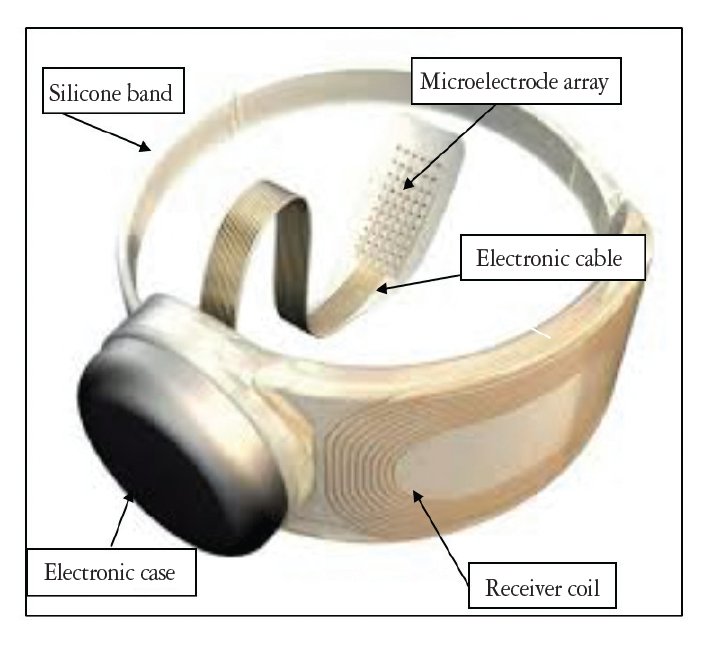
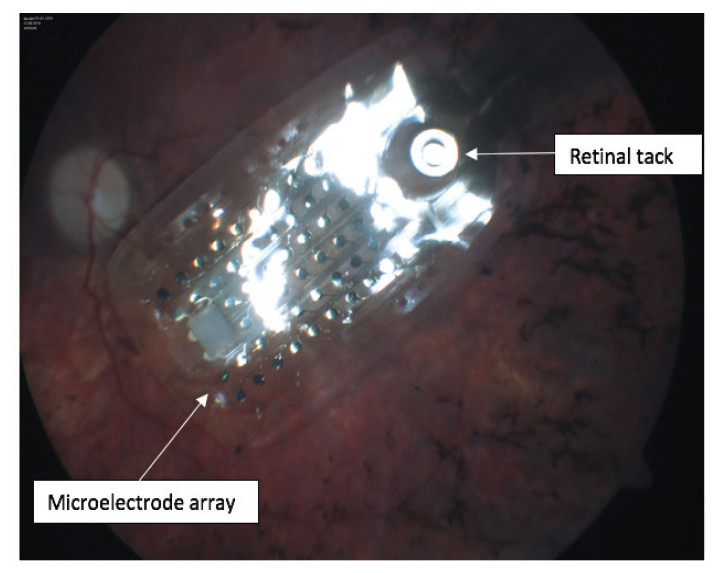
In outer retinal degenerative diseases such as retinitis pigmentosa, choroideremia, and geographic atrophy, 30% of the ganglion cell layer in the macula remains intact. With subretinal and epiretinal prostheses, these inner retinal cells are stimulated with controlled electrical current by either a microphotodiode placed in the subretinal area or a microelectrode array tacked to the epiretinal region. As the patient learns to interpret the resulting phosphene patterns created in the brain through special rehabilitation exercises, their orientation, mobility, and quality of life increase. Implants that stimulate the lateral geniculate nucleus or visual cortex are currently being studied for diseases in which the ganglion cells and optic nerve are completely destroyed.
Keywords: Artificial vision, bionic eye, visual prosthesis, Argus II, retinal prosthesis, outer retinal degeneration, retinitis pigmentosa, phosphene
Introduction
Nearly half of visual impairment worldwide is caused by retinal diseases. Degenerative retinal diseases such as retinitis pigmentosa (RP), choroideremia, and age-related macular degeneration (AMD) begin in the outer retinal layers and progress gradually, with the inner retinal layers remaining largely unaffected until advanced stages of disease. Histopathological studies have shown that 70% of photoreceptors are lost in AMD, while 93% of the retinal ganglion cells (RGCs) survive. RP mainly affects the photoreceptor layer; in the macular region, 78-88% of the inner nuclear layer (INL) and approximately 30% of the ganglion cell layer (GCL) remains intact. RP leads to cell loss in all retinal layers in the extramacular region; the INL and GCL are relatively less preserved than the macular region. There is no difference between the different genetic types of RP in terms of macular cell loss. In the extramacular region, more cells are preserved in autosomal-dominant RP.,,
Neovascular AMD is treated at significant rates using intravitreal anti-VEGF drug injections, but there is not yet a proven effective treatment for geographic atrophy (GA), an advanced stage of dry AMD. RPE65 gene therapy and slow-release ciliary neurotrophic factor implants are being used in the treatment of RP, and stem cell research is ongoing. However, there is no proven, definite treatment approach. Retinal prostheses developed in recent years are promising for eyes with severe visual impairment due to outer retinal degeneration.,
Conclusion
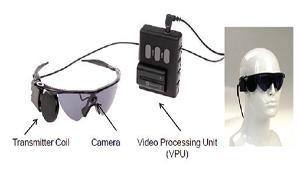
Studies on the Argus II epiretinal prosthesis performed to date have demonstrated the long-term safety and potential benefits of controlled chronic electrical stimulation in patients with advanced visual impairment due to outer retinal degeneration associated with conditions such as RP, choroideremia, and GA. Some of the missing pieces in information obtained through artificial vision are filled in by the brain based on previous experiences. The limited number of clinical studies performed with various other retinal prostheses other than the Argus II system have also yielded promising results. However, each type of prosthesis has its own advantages and disadvantages. Cortical implants, which are currently in the preclinical study phase, may provide artificial vision to patients with complete retina and optic nerve destruction.
References
Kien TT, Maul T, Bargiela A. A review of retinal prosthesis approaches. Int J Mod Phys. 2012;9:209-231.
Rachitskaya AV, Yuan A. Argus II retinal prosthesis system: An update. Ophthalmic Genet. 2016;37:260-266.
Falabella P, Nazari H, Schor P. Argus II retinal prosthesis system. In: Gabel VP ed. Artificial Vision: A Clinical Guide. Springer, 2017:49-63.
Chuang AT, Margo CE, Greenberg PB. Retinal implants: a systematic review. Br J Ophthalmol. 2014;98:852-856.
Asher A, Segal WA, Baccus SA, Yaroslavsky LP, Palanker DV. Image processing for a high-resolution optoelectronic retinal prosthesis. IEEE Trans Biomed Eng. 2007;54:993-1004.
ORION Cortical visual prosthesis. Second Sight Medical Products, Sylmar, CA-USA.
Khanna VK. Retinal Prostheses. Implantable Medical Electronics. Springer International Publishing Switzerland: 2016:393-408.
Rao C, Yuan X-H, Zhang SJ, Wang QL, Huang YS. Epiretinal prosthesis for outer retinal degenerative diseases. Int J Ophthalmol. 2008;1:273-276.
Coussa RG, Traboulsi EI. Choroideremia: a review of general findings and pathogenesis. Ophthalmic Genet. 2012;33:57-65.
Luo YH, da Cruz L. The Argus II retinal prosthesis system. Prog Retin Eye Res. 2016;50:89-107.
Argus II retinal prosthesis system-H110002. Last updated 13 Jan 2015. http://www.fda.gov/medicaldevices/productsandmedicalprocedures/deviceapprovalsandclearances/recently-approveddevices/ucm343162.htm.Accessed 7 Feb 2015
Verlag AG. Retina Implant. In: Walter P, ed. Care-Basics, Procedures and Follow-up UNI-MED Bremen, London, Boston; 2014.
Argus II Retinal Prosthesis System: Surgical Manual, Second Sight Medical Products, Inc. 2014.
Ho AC, Humayun MS, Dorn JD, da Cruz L, Dagnelie G, Handa J, Barale PO, Sahel JA, Stanga PE, Hafezi F, Safran AB, Salzmann J, Santos A, Birch D, Spencer R, Cideciyan AV, de Juan E, Duncan JL, Eliott D, Fawzi A, Olmos de Koo LC, Brown GC, Haller JA, Regillo CD, Del Priore LV, Arditi A, Geruschat DR, Greenberg RJ; Argus II Study Group. Long-term results from an epiretinal prosthesis to restore sight to the blind. Ophthalmology. 2015;122:1547-1554.
Stanga P. Implantation of Argus II in patients with advanced dry AMD. Manchester Eye Hospital, U.K. https://clinicaltrials.gov/ct2/show/NCT02227498
Retinal Prosthesis: A Clinical Guide to Successful Implementation. Eds: Humayun MS, de Koo LCO, Springer; 2018.
Humayun MS, Dorn JD, da Cruz L, Dagnelie G, Sahel JA, Stanga PE, Cideciyan AV, Duncan JL, Eliott D, Filley E, Ho AC, Santos A, Safran AB, Arditi A, Del Priore LV, Greenberg RJ; Argus II Study Group. Interim results from the international trial of Second Sight’s visual prosthesis. Ophthalmology. 2012;119:779-788.
Rizzo S, Belting C, Cinelli L, Allegrini L, Genovesi-Ebert F, Barca F, di Bartolo E. The Argus II Retinal Prosthesis: 12-month outcomes from a single-study center. Am J Ophthalmol. 2014;157:1282-1290.
Özmert E, Demirel S. Endoscope-assisted and controlled Argus II epiretinal prosthesis implantation in late-stage retinitis pigmentosa: A report of 2 cases. Case Rep Ophthalmol. 2016;7:315-324.
Özmert E, Baskak B, Arslantaş R, Kır Y, Kuşman A, Baran Z. Measurement of visual cortex activity with near-infrared spectroscopy after endoscope assisted Argus II epiretinal prosthesis implantation in retinitis pigmentosa patients. Artificial Vision, The International Symposium on Visual Prosthetics, Aachen University - Germany; 2017:14.
Scarapicchia V, Brown C, Mayo C, Gawryluk JR. Functional magnetic resonance imaging and functional near-infrared spectroscopy: insight from combined recording studies. Front Hum Neurosci. 2017;11:419.
Humayun MS, Dorn JD, Ahuja AK, Caspi A, Filley E, Dagnelie G, Salzmann J, Santos A, Duncan J, daCruz L, Mohand-Said S, Eliott D, McMahon MJ, Greenberg RJ. Preliminary 6 month results from the Argus II epiretinal prosthesis feasibility study. Conf Proc IEEE Eng Med Biol Soc. 2009;4566-4568.
Ahuja AK, Yeoh J, Dorn JD, Caspi A, Wuyyuru V, McMahon MJ, Humayun MS, Greenberg RJ, Dacruz L. Factors affecting perceptual threshold in Argus II retinal prosthesis subjects. Transl Vis Sci Technol. 2013;2:1.
Sahel J, Mohand-Said S, Stanga P, Caspi A, Greenberg R,. Acuboost: enhancing the maximum acuity of the Argus II retinal prosthesis system. Invest Ophthalmol Vis Sci. 2013;54:1389.
Stanga P, Sahel J, Mohand-Said S, daCruz L, Capi A, Francesco M, Greenberg R; Argus II Study Group. Face detection using the Argus II retinal prosthesis system. Invest Ophthalmol Vis Sci. 2013;54:1766.

