

Abstract
Avascular peripheral retina in an infant is a common characteristic of numerous pediatric retinal vascular disorders and often presents a diagnostic challenge to the clinician. In this review, key features of each disease in the differential diagnosis, from retinopathy of prematurity, familial exudative vitreoretinopathy, Coats disease, incontinentia pigmenti, Norrie disease, and persistent fetal vasculature, to other rare hematologic conditions and telomere disorders, will be discussed by expert ophthalmologists in the field.
Keywords: Avascular retina, retinopathy of prematurity, familial exudative vitreoretinopathy, Coats disease, incontinentia pigmenti, persistent fetal vasculature, Norrie disease
Introduction
Pediatric retinal vascular diseases are the leading causes of childhood blindness throughout the world. Therefore, correct and timely diagnosis of an infant presenting with a retinal vascular disease is of paramount importance. However, this can often be challenging.
Avascular peripheral retina is a common feature of many pediatric retinal vascular diseases. Therefore, the clinician should seek out key features that will help differentiate the various diseases that may present in such a manner. Clinical evaluation should begin with a detailed medical history, including gestational age, weight, birth history, neonatal course, and the presence of any systemic findings. Family history, including known ocular disease in the immediate family or close relatives and the presence of parental consanguinity should be questioned. A comprehensive ophthalmological examination should then be performed. Also, examination of available family members often provides important clues for an accurate diagnosis.
In this article, experienced ophthalmologists in the field will review key points of diseases included in the differential diagnosis of a pediatric patient presenting with avascular peripheral retina. The major differential diagnosis includes retinopathy of prematurity (ROP), familial exudative vitreoretinopathy (FEVR), Coats’ disease, incontinentia pigmenti (IP), Norrie disease, and persistent fetal vasculature (PFV). In addition, other rare conditions that may present with avascular peripheral retina, such as hematologic and inflammatory conditions and telomere disorders, will also be discussed.
Conclusions
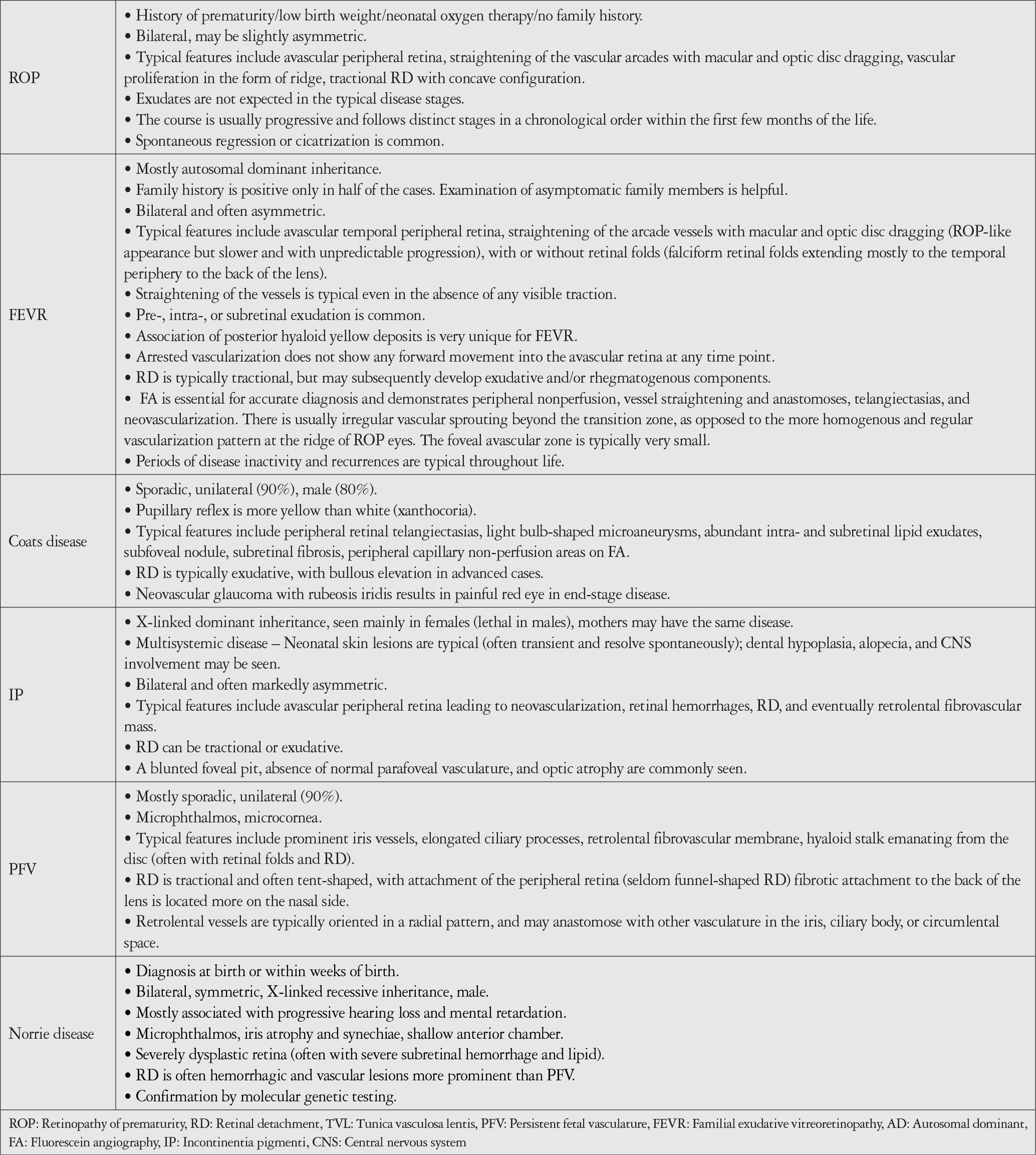
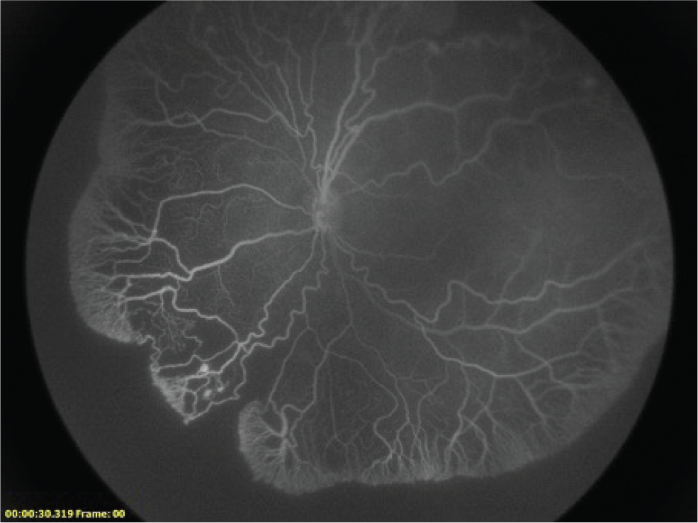
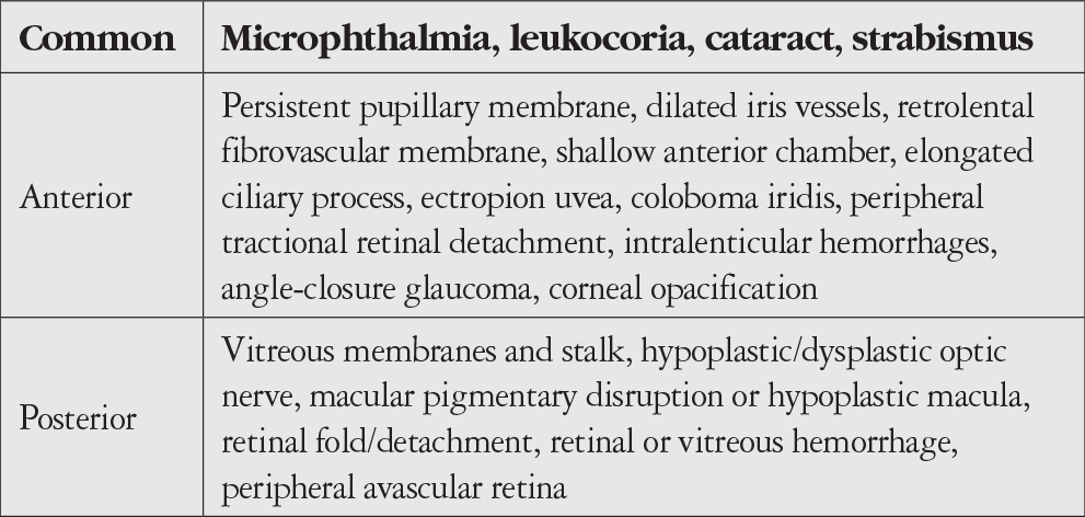
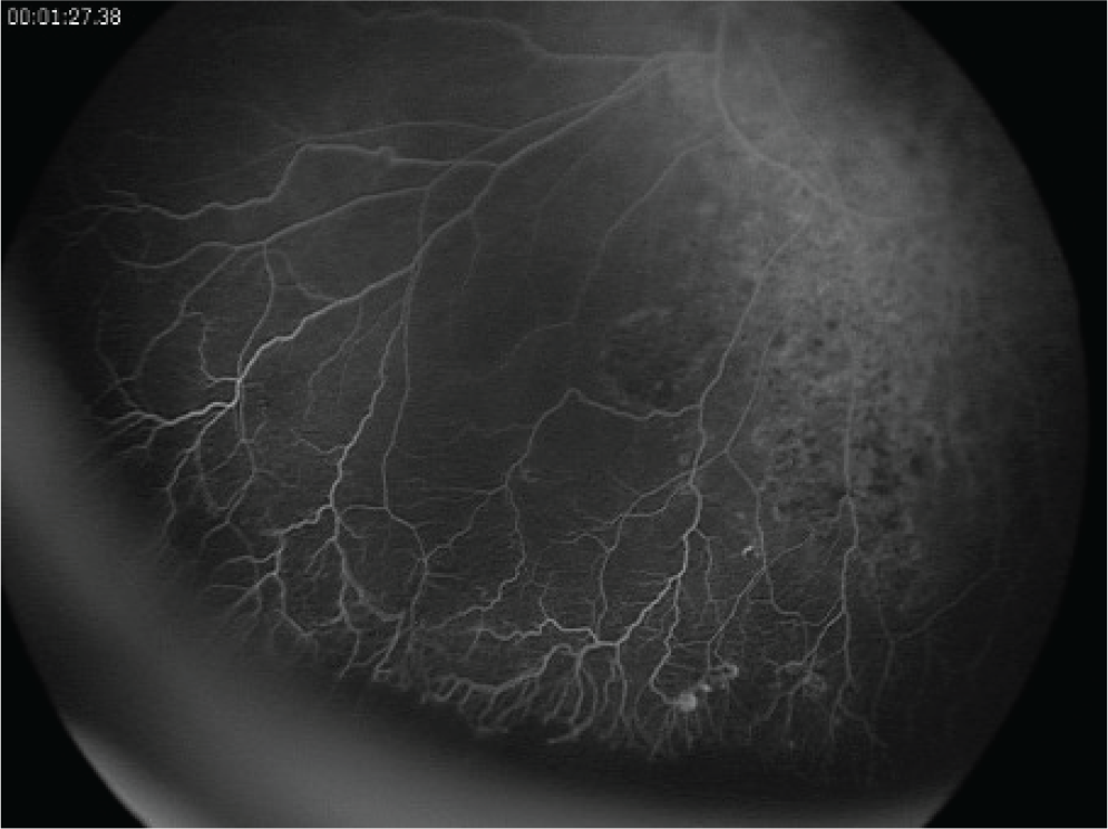
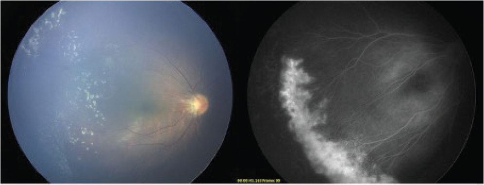
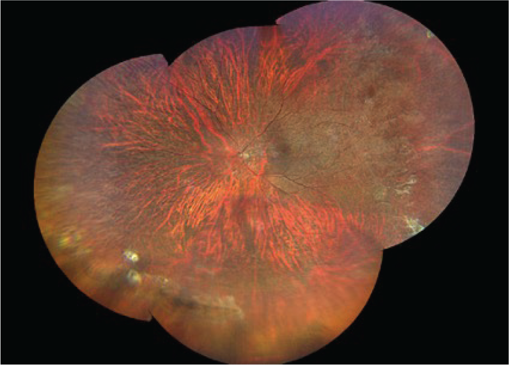
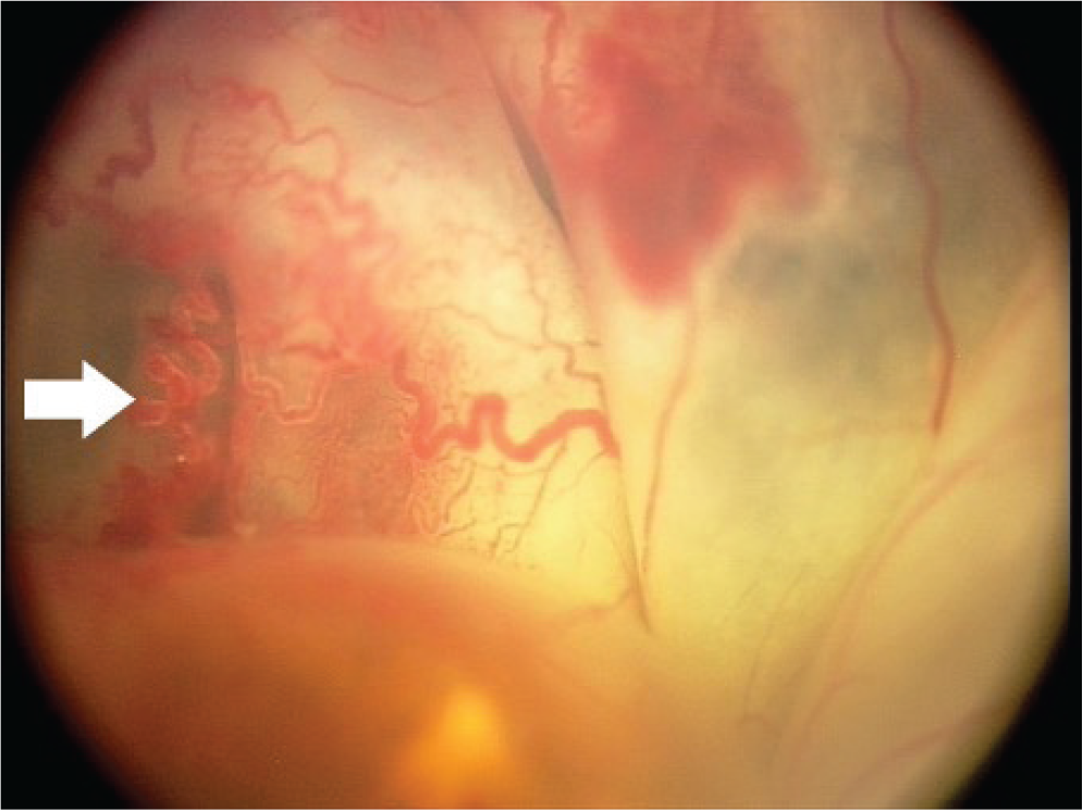
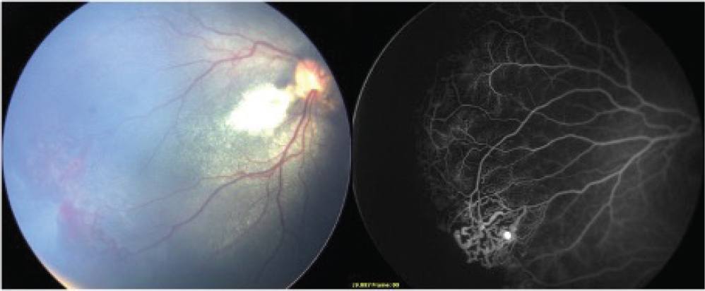
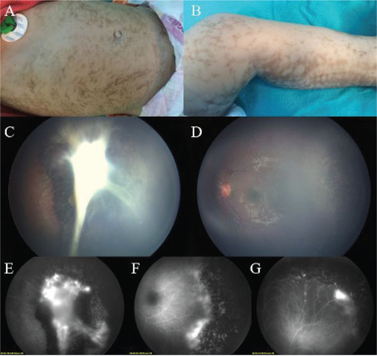
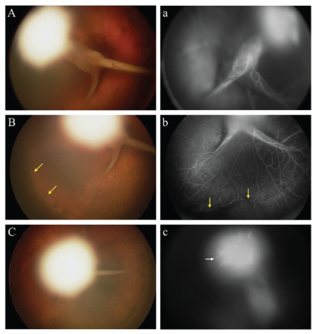
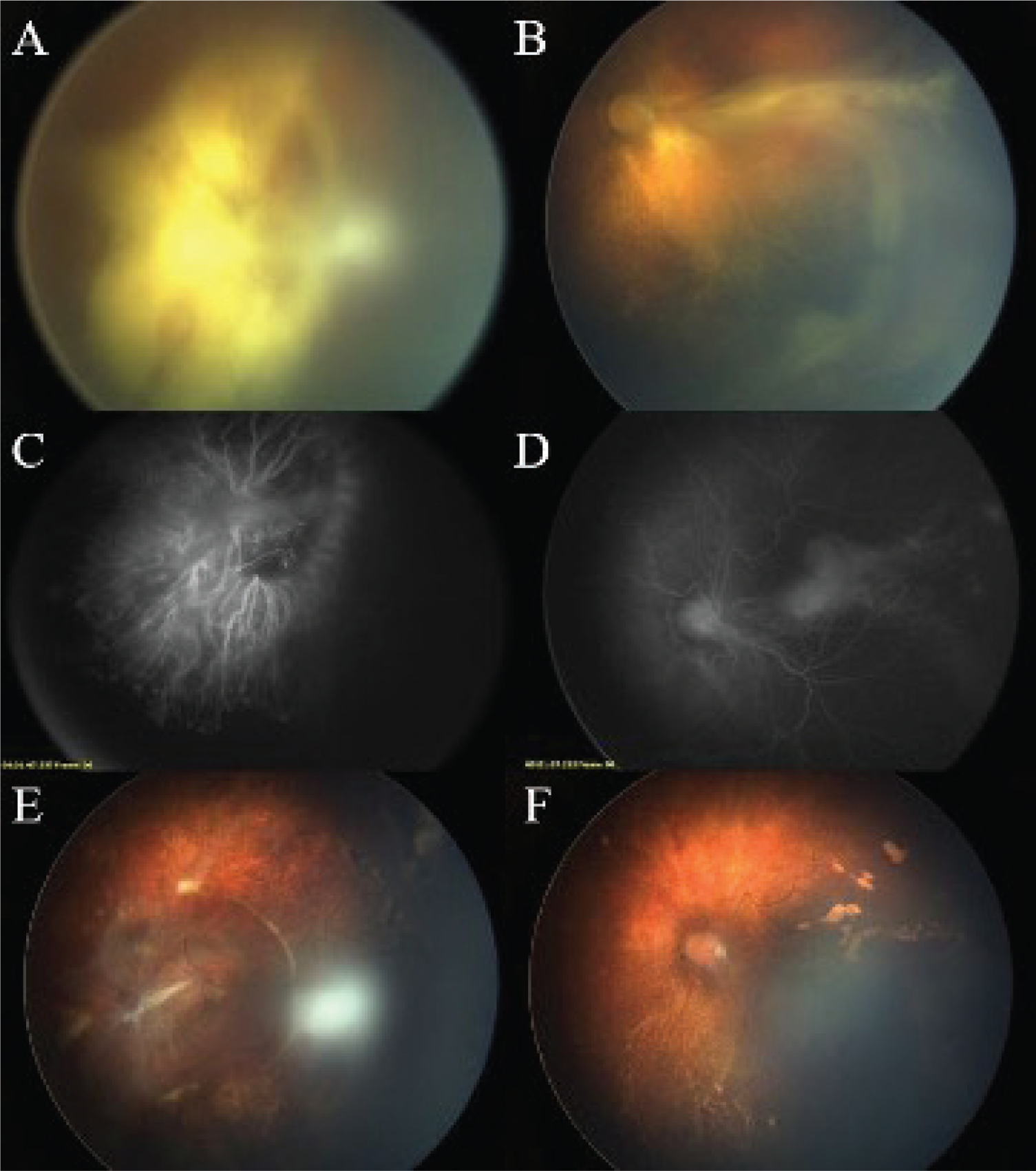
To summarize, the most common cause of avascular peripheral retina is ROP. Although ophthalmoscopic features may overlap with other retinal vascular diseases, patients who develop ROP almost uniformly have prematurity and low birth weight. Furthermore, active neovascularization in late infancy, significant asymmetry, and family history of disease are not expected. In term babies or those with marked asymmetry, accompanying systemic findings, or a history of similar disease in close relatives, less common causes of similar clinical presentation should be considered in the differential diagnosis. summarizes the key differentiating features of these causes.
References
Spandau U, Kim SJ. Pediatric Retinal Vascular Diseases. 2019. doi:10.1007/978-3-030-13701-4
Michaelson IC. The mode of development of the vascular system of the retina with some observations on its significance for certain retinal diseases. Trans Ophthalmol Soc UK. 1948;68:137-180.
Flower RW, McLeod DS, Lutty GA, Goldberg B, Wajer SD. Postnatal retinal vascular development of the puppy. Invest Ophthalmol Vis Sci. 1985;26:957-968.
Smith LEH. Pathogenesis of retinopathy of prematurity. Semin Neonatol. 2003;8:469-473.
Chan-Ling T. Vasculogenesis and Angiogenesis in Formation of the Human Retinal Vasculature. In: Penn JS, ed. Retinal and Choroidal Angiogenesis. 1st ed. Springer; 2008:119-138. doi:10.1007/978-1-4020-6780-8_6
Hughes S, Yang H, Chan-Ling T. Vascularization of the human fetal retina: Roles of vasculogenesis and angiogenesis. Invest Ophthalmol Vis Sci. 2000;41:1217-1228.
Lepore D, Ji MH, Quinn GE, Amorelli GM, Orazi L, Ricci D, Mercuri E. Functional and Morphologic Findings at Four Years After Intravitreal Bevacizumab or Laser for Type 1 ROP. Ophthalmic Surg Lasers Imaging Retina. 2020;51:180-186.
Chiang MF, Quinn GE, Fielder AR, Ostmo SR, Paul Chan RV, Berrocal A, Binenbaum G, Blair M, Peter Campbell J, Capone A Jr, Chen Y, Dai S, Ells A, Fleck BW, Good WV, Elizabeth Hartnett M, Holmstrom G, Kusaka S, Kychenthal A, Lepore D, Lorenz B, Martinez-Castellanos MA, Özdek Ş, Ademola-Popoola D, Reynolds JD, Shah PK, Shapiro M, Stahl A, Toth C, Vinekar A, Visser L, Wallace DK, Wu WC, Zhao P, Zin A. International Classification of Retinopathy of Prematurity, Third Edition. Ophthalmology. 2021;128:51-68.
Gilmour DF. Familial exudative vitreoretinopathy and related retinopathies. Eye (Lond). 2015;29:1-14.
Ranchod TM, Ho LY, Drenser KA, Capone A Jr, Trese MT. Clinical presentation of familial exudative vitreoretinopathy. Ophthalmology. 2011;118:2070-2075.
John VJ, McClintic JI, Hess DJ, Berrocal AM. Retinopathy of Prematurity Versus Familial Exudative Vitreoretinopathy: Report on Clinical and Angiographic Findings. Ophthalmic Surg Lasers Imaging Retina. 2016;47:14-19.
Musada GR, Syed H, Jalali S, Chakrabarti S, Kaur I. Mutation spectrum of the FZD-4, TSPAN12 AND ZNF408 genes in Indian FEVR patients. BMC Ophthalmol. 2016;16:90.
Pendergast SD, Trese MT. Familial exudative vitreoretinopathy: Results of surgical management. Ophthalmology. 1998;105:1015-1023.
Kashani AH, Learned D, Nudleman E, Drenser KA, Capone A, Trese MT. High prevalence of peripheral retinal vascular anomalies in family members of patients with familial exudative vitreoretinopathy. Ophthalmology. 2014;121:262-268.
Shukla D, Singh J, Sudheer G, Soman M, John RK, Ramasamy K, Perumalsamy N. Familial exudative vitreoretinopathy (FEVR). Clinical profile and management. Indian J Ophthalmol. 2003;51:323-328.
Shields JA, Shields CL, Honavar SG, Demirci H. Clinical variations and complications of Coats disease in 150 cases: the 2000 Sanford Gifford Memorial Lecture Am J Ophthalmol. 2001;131:561-571.
Shields CL, Udyaver S, Dalvin LA, Lim LS, Atalay HT, L Khoo CT, Mazloumi M, Shields JA. Coats disease in 351 eyes: Analysis of features and outcomes over 45 years (by decade) at a single center. Indian J Ophthalmol. 2019;67:772-783.
B Blair MP, Ulrich JN, Elizabeth Hartnett M, Shapiro MJ. Peripheral retinal nonperfusion in fellow eyes in coats disease. Retina. 2013;33:1694-1699.
Kusaka S. Surgical management of coats disease. Asia Pac J Ophthalmol (Phila). 2018;7:156-159.
Ucgul AY, Ozdek S, Ertop M, Atalay HT. External Drainage Alone Versus External Drainage With Vitrectomy in Advanced Coats Disease. Am J Ophthalmol. 2021;222:6-14.
He YG, Wang H, Zhao B, Lee J, Bahl D, McCluskey J. Elevated vascular endothelial growth factor level in Coats’ disease and possible therapeutic role of bevacizumab. Graefes Arch Clin Exp Ophthalmol. 2010;248:1519-1521.
Mano F, Matsushita I, Kondo H, Utamura S, Kondo C, Kusaka S. Vitrectomy and external drainage of subretinal fluid containing high concentration of vascular endothelial growth factor for advanced coats disease. Sci Rep. 2021;11:19333.
Cai X, Zhao P, Zhang Q, Jin H. Treatment of stage 3 Coats’ disease by endolaser photocoagulation via a two-port pars plana nonvitrectomy approach. Graefe’s Arch Clin Exp Ophthalmol. 2015;253:999-1004.
Li AS, Capone A Jr, Trese MT, Sears JE, Kychenthal A, De la Huerta I, Ferrone PJ. Long-Term Outcomes of Total Exudative Retinal Detachments in Stage 3B Coats Disease. Ophthalmology. 2018;125:887-893.
Marques GF, Tonello CS, Sousa JM. Incontinentia pigmenti or Bloch-Sulzberger syndrome: a rare X-linked genodermatosis. An Bras Dermatol. 2014;89:486-489.
Ferrone PJ, Awner S. Proliferative retinopathies in children. in Pediatric Retina (2011). doi:10.1007/978-3-642-12041-1_15.
Lee TC, Chiang MF. Pediatric retinal vascular diseases. Retina 5th ed. 2012. doi:10.1016/B978-1-4557-0737-9.00061-8.
Buinauskiene J, Buinauskaite E, Valiukeviciene S. Incontinentia pigmenti (Bloch-Sulzberger syndrome) in neonates. Medicina (Kaunas). 2005;41:496-499.
Pereira MA, Mesquita LA, Budel AR, Cabral CS, Feltrim Ade S. X-linked incontinentia pigmenti or Bloch-Sulzberger syndrome: a case report. An Bras Dermatol. 2010;85:372-375.
Minić S, Trpinac D, Gabriel H, Gencik M, Obradović M. Dental and oral anomalies in incontinentia pigmenti: a systematic review. Clin Oral Investig. 2013;17:1-8.
Cammarata-Scalisi F, Fusco F, Ursini MV. Incontinentia Pigmenti. Actas Dermosifiliogr (Engl Ed). 2019;110:273-278.
Minić S, Trpinac D, Obradović M. Systematic review of central nervous system anomalies in incontinentia pigmenti. Orphanet J Rare Dis. 2013;8:25.
Peng J, Zhang Q, Long X, Zhang J, Huang Q, Li Y, She K, Zhao P. Incontinentia pigmenti-associated ocular anomalies of paediatric incontinentia pigmenti patients in China. Acta Ophthalmol. 2019;97:265-272.
Narang S, Sindhu M, Jain S, Jinagal J. Retinal imaging in incontinentia pigmenti. Indian J Ophthalmol. 2019;67:944-945.
K Kim SJ, Yang J, Liu G, Huang D, Campbell JP. Optical Coherence Tomography Angiography and Ultra-Widefield Optical Coherence Tomography in a Child With Incontinentia Pigmenti. Ophthalmic Surg Lasers Imaging Retina. 2018;49:273-275.
Sen A, Shenoy P, Mitra A, Jain T. Multimodal retinal imaging of a 6-year-old male child with incontinentia pigmenti. Indian J Ophthalmol. 2019;67:942-943.
Martínez-Gayosso A, García-Romero MT. Incontinentia pigmenti: multisistemic genodermatosis. Bol Med Hosp Infant Mex. 2020;77:112-118.
Chen C, Xiao H, Ding X. Persistent fetal vasculature. Asia Pac J Ophthalmol (Phila) 2019;8:86-95.
Prakhunhungsit S, Berrocal AM. Diagnostic and Management Strategies in Patients with Persistent Fetal Vasculature: Current Insights. Clin Ophthalmol. 2020;14:4325-4335.
Jeng-Miller KW, Joseph A, Baumal CR. Fluorescein Angiography in Persistent Fetal Vasculature. Ophthalmology. 2017;124:455.
Laura DM, Staropoli PC, Patel NA, Yannuzzi NA, Nolan RP, Al-Khersan H, Fan KC, Flynn HW Jr, Acon D, Negron CI, Berrocal AM. Widefield Fluorescein Angiography in the Fellow Eyes of Patients with Presumed Unilateral Persistent Fetal Vasculature. Ophthalmol Retina. 2021;5:301-307.
Shen JH, Liu L, Wang NK, Hwang YS, Chen KJ, Chao AN, Lai CC, Chen TL, Wu WC. Fluorescein Angiography Findings In Unilateral Persistent Fetal Vasculature. Retina. 2020;40:572-580.
Peripheral vascular abnormalities detected by fluorescein angiography in contralateral eyes of patients with persistent fetal vasculature. Ophthalmic Surg. 2020;51:506-511.
Staropoli PC, Yannuzzi NA, Patel NA, Negron CI, Prakhunhungsit S, Berrocal AM. Novel Frizzled-4 Mutation Is Associated With Familial Exudative Vitreoretinopathy Mimicking Persistent Fetal Vasculature. J Pediatr Ophthalmol Strabismus 2020;57:4-7.
Sisk RA, Berrocal AM, Feuer WJ, Murray TG. Visual and anatomic outcomes with or without surgery in persistent fetal vasculature. Ophthalmology. 2010;117:2178-2183.
Zahavi A, Weinberger D, Snir M, Ron Y. Management of severe persistent fetal vasculature: case series and review of the literature. Int Ophthalmol. 2019;39:579-587.
Ozdemir Zeydanli, E. & Ozdek, S. Surgical Results of Posterior Persistent Vasculature Syndrome. in Turkish Ophthalmological Association Virtual National Congress and Live Surgery Compound Meeting; (2020).
Bosjolie A, Ferrone P. Visual outcome in early vitrectomy for posterior persistent fetal vasculature associated with traction retinal detachment. Retina. 2015;35:570-576.
Sims KB. NDP-Related Retinopathies. GeneReviews; 1993.
Warburg M. Norrie’s disease. A congenital progressive oculo-acoustico-cerebral degeneration. Acta Ophthalmol(Copenh). 1966;(Suppl 89):1-47.
Warburg M. Norrie’s disease--differential diagnosis and treatment. Acta Ophthalmol (Copenh). 1975;53:217-236.
Walsh MK, Drenser KA, Capone A Jr, Trese MT. Early vitrectomy effective for Norrie disease. Arch Ophthalmol. 2010;128:456-460.
Spandau UHM, Kim SJ. Pediatric Retinal Vascular Diseases. Springer International Publishing; 2019:15-18. doi:10.1007/978-3-030-13701-4_2
Duke JR. Pseudoglioma in children: Aspects of clinical and pathological diagnosis. South Med J. 1958;51:754-759.
Dickinson JL, Sale MM, Passmore A, FitzGerald LM, Wheatley CM, Burdon KP, Craig JE, Tengtrisorn S, Carden SM, Maclean H, Mackey DA. Mutations in the NDP gene: contribution to Norrie disease, familial exudative vitreoretinopathy and retinopathy of prematurity. Clin Exp Ophthalmol. 2006;34:682-688.
Walsh MK, Drenser KA, Capone A Jr, Trese MT. Norrie disease vs familial exudative vitreoretinopathy. Arch Ophthalmol. 2011;129:819-820.
Walsh MK, Drenser KA, Capone A Jr, Trese MT. Early vitrectomy effective for bilateral combined anterior and posterior persistent fetal vasculature syndrome. Retina. 2010;30(4 Suppl):2-8.
Smith SE, Mullen TE, Graham D, Sims KB, Rehm HL. Norrie disease: extraocular clinical manifestations in 56 patients. Am J Med Genet A. 2012;158:1909-1917.
Halpin C, Owen G, Gutiérrez-Espeleta GA, Sims K, Rehm HL. Audiologic features of Norrie disease. Ann Otol Rhinol Laryngol. 2005;114:533-538.
Marlar RA, Montgomery RR, Broekmans AW. Report on the Diagnosis and Treatment of Homozygous Protein C Deficiency. Thromb Haemost. 1989;61:529-531.
Ghassemi F, Abdi F, Esfahani M. Ophthalmic manifestations of congenital protein C deficiency: a case report and mini review. BMC Ophthalmol. 2020;20:282.
T Taher NO, Ghaddaf AA, Al-Ghamdi SA, Homsi JJ, Al-Harbi BJ, Alomari LK, Almarzouki HS. Intravitreal Anti-vascular Endothelial Growth Factor Injection for Retinopathy of Prematurity: A Systematic Review and Meta-Analysis. Front Med (Lausanne). 2022;9:884608.
Adams FH, Oliver CP. Hereditary deformities in man: Due to arrested development. J Hered. 1945;36:3-7.
Lehman A, Stittrich AB, Glusman G, Zong Z, Li H, Eydoux P, Senger C, Lyons C, Roach JC, Patel M. Diffuse angiopathy in Adams-Oliver syndrome associated with truncating DOCK6 mutations. Am J Med Genet A. 2014;164:2656-2662.
Tan W, Palmby TR, Gavard J, Amornphimoltham P, Zheng Y, Gutkind JS. An essential role for Rac1 in endothelial cell function and vascular development. FASEB J. 2008;22:1829-1839.
Peralta-Calvo J, Pastora N, Casa-Ventura YG, Hernandez-Serrano R, Abelairas J. Peripheral ischemic retinopathy in Adams-Oliver syndrome. Arch Ophthalmol. 2012;130:1078-1080.
Revesz T, Fletcher S, al-Gazali LI, DeBuse P. Bilateral retinopathy, aplastic anaemia, and central nervous system abnormalities: a new syndrome? J Med Genet. 1992;29:673-675.
Gupta MP, Talcott KE, Kim DY, Agarwal S, Mukai S. Retinal findings and a novel TINF2 mutation in Revesz syndrome: Clinical and molecular correlations with pediatric retinal vasculopathies. Ophthalmic Genet. 2017;38:51-60.
McElnea EM, van der Spek N, Smith O, Fitzsimon S, Patel CK, O’Marcaigh A. Revesz syndrome masquerading as bilateral cicatricial retinopathy of prematurity. J AAPOS. 2013;17:634-636.
Asano M, Tsukamoto S, Sonoda KH, Kondo H. Revesz syndrome with bilateral retinal detachments successfully treated by pars plana vitrectomy. Am J Ophthalmol Case Rep. 2021;23:101137.
Riyaz A, Riyaz N, Jayakrishnan MP, Mohamed Shiras PT, Ajith Kumar VT, Ajith BS. Revesz syndrome. Indian J Pediatr. 2007;74:862-863.

