

Abstract
Objectives:
To retest the performance of Pentacam parameters in the detection of eyes with subclinical keratoconus (KC) and mild KC based on different definitions from the Amsler-Krumeich (AK), Collaborative Longitudinal Evaluation of Keratoconus (CLEK), and ABCD systems.
Materials and Methods:
This cross-sectional university-based study comprised 24 eyes with subclinical KC, 144 eyes with mild KC (based on AK in 101 eyes, CLEK in 28 eyes, and ABCD in 15 eyes), and 70 controls. Diagnostic ability of the thinnest point (TP) pachymetry, KISA% index, inferior-superior asymmetry, corneal aberrations, Pentacam indices, front/back elevations, pachymetric progression index, Ambrósio-Relational Thickness (ARTmax), and Belin/Ambrósio Enhanced Ectasia Display scores (Df, Db, Dp, Dt, Da, and D-final) were evaluated.
Results:
ARTmax (83.3% sensitivity/74.3% specificity) had the highest ability in distinguishing subclinical KC from normal, followed by TP pachymetry, Dt, and Da. D-final showed excellent sensitivity/specificity in mild KC diagnosis based on AK (98%/100%) and CLEK (97.4%/100%) descriptions. In the mild KC-ABCD group, index of vertical asymmetry accurately detected all eyes with mild KC and 97.1% of the controls.
Conclusion:
This study points out the gray zone in the detection of eyes with subclinical and mild KC due to overlapping terminology and grading criteria. Pentacam parameters seem to have modest capability in subclinical KC detection, indicating the necessity for additional diagnostic modalities. However, eyes with mild KC can be diagnosed with high accuracy using Pentacam parameters, although the strongest parameters may vary according to the definition of “mild KC.” Nevertheless, uniform and definitive criteria for subclinical and clinical KC classification are required for a diagnostic and therapeutic consensus in KC.
Keywords: Diagnosis, Pentacam, Scheimpflug, subclinical keratoconus
Introduction
Keratoconus (KC) is an asymmetrically bilateral progressive corneal ectasia characterized by visual deterioration and stromal thinning. In moderate and advanced stages, the diagnosis of KC can easily be made based on apparent clinical and topographical findings, whereas detecting eyes with KC in its earliest form remains a challenge.
There is growing interest in developing a powerful parameter or formula for distinguishing subclinical cases of KC to avoid iatrogenic post-laser ectasia.,,,,, However, there is no consensus in the literature on the nomenclature for early stages of KC.,,,,,
A recent systematic review reported that the most commonly used definition of subclinical KC is an eye with topographic signs of KC and/or suspicious topographic findings with normal slit-lamp examination and KC in the fellow eye.,,,,, Regarding clinical KC, various classification systems such as the Amsler-Krumeich (AK), KC severity score, Collaborative Longitudinal Evaluation of Keratoconus (CLEK), ABCD, and RETICS (Red Temática de Investigación Cooperativa en Salud) were introduced to grade disease severity.,,,, The RETICS was developed by Alió et al. as a visual limitation-based KC classification.
Recently, Belin and Duncan proposed the ABCD system, which incorporates anterior/posterior corneal curvature data, thinnest point (TP) on pachymetry, and corrected distance visual acuity (CDVA) for progression follow-up in eyes with KC. The ABCD system has been also integrated into the Pentacam HR software (Oculus Optikgerate GmBH, Wetzlar, Germany).
Pentacam technology has an important place in the diagnosis of KC because it provides a variety of quantitative parameters, and the utility of these parameters in early KC detection is still being tested. However, previous studies reported diverse sensitivity and specificity values for Pentacam parameters in the diagnosis of subclinical and mild KC due to overlaps among these definitions and a lack of globally accepted uniform criteria.,,,,,,,,
This study aimed to re-evaluate the performance of Pentacam parameters in the discrimination of eyes with subclinical KC and mild KC using different definitions of “mild KC” from the AK, CLEK, and Belin ABCD systems against the backdrop of previously published similar studies.
Materials and Methods
The Pamukkale University Non-Interventional Clinical Research Ethics Committee approved the study protocol (decision no: 23, date: 08.12.2020) and the tenets of Declaration of Helsinki on the use of human subjects in research were followed. This retrospective university-based, single-center, cross-sectional study included 24 eyes with subclinical KC (24 patients), 144 eyes with mild KC (144 patients), and 70 control eyes with normal tomography (70 subjects).
All included subjects had reliable records for ophthalmological examinations including CDVA (Snellen) measurement, slit-lamp biomicroscopic examination, dilated fundus examination, and Pentacam imaging. Contact lens wearers were requested to remove their contact lenses prior to the measurements (at least 2 weeks for soft contact lenses and 3 weeks for hard contact lenses).
Each patient was only included in one group to ensure independence of the groups. One eye per patient was used for the statistical analysis. The randomization function of SPSS Statistics for Windows version 24.0 (IBM Corp., Armonk, NY, USA) was used for eye selection in bilateral cases.
Results
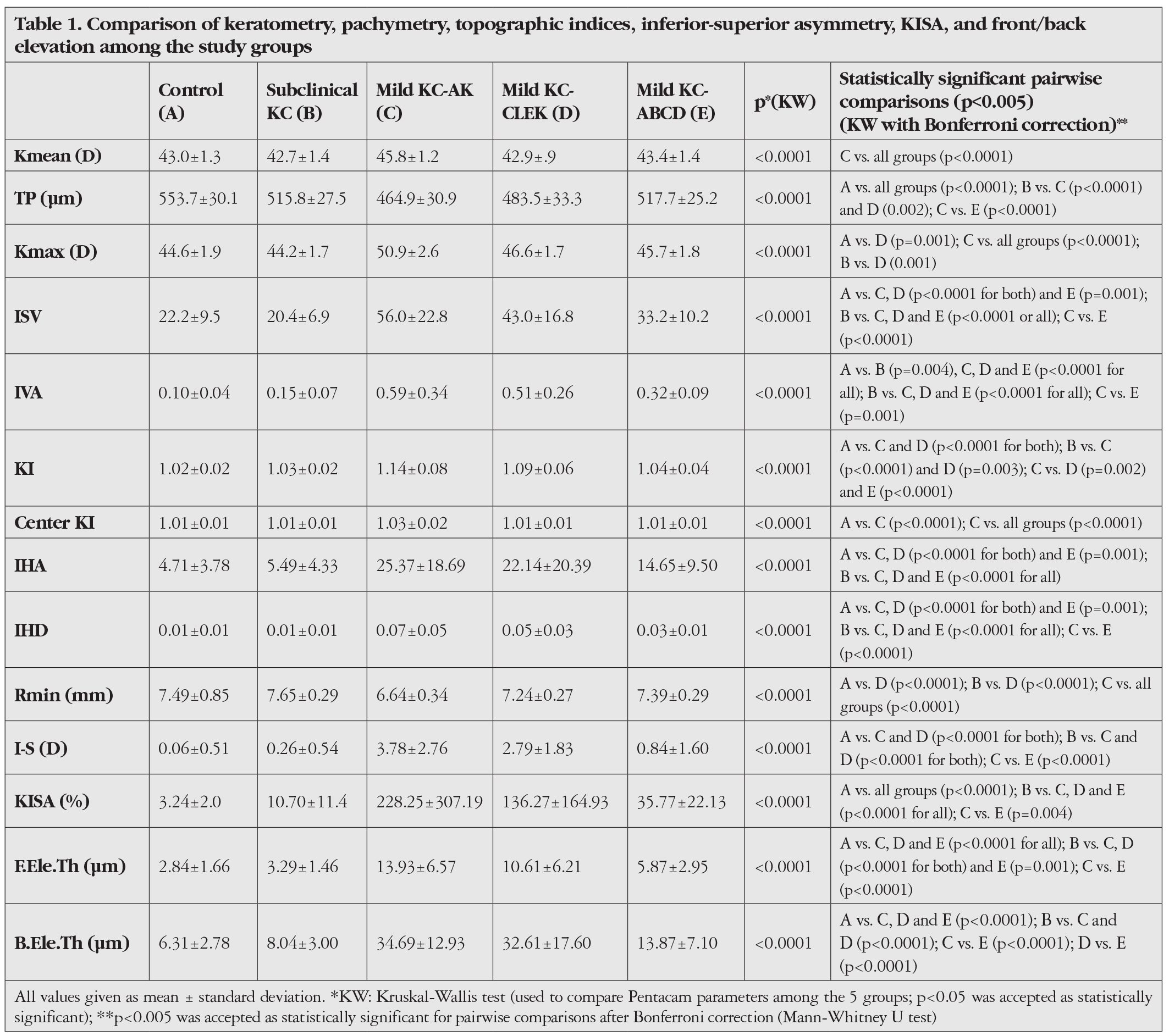
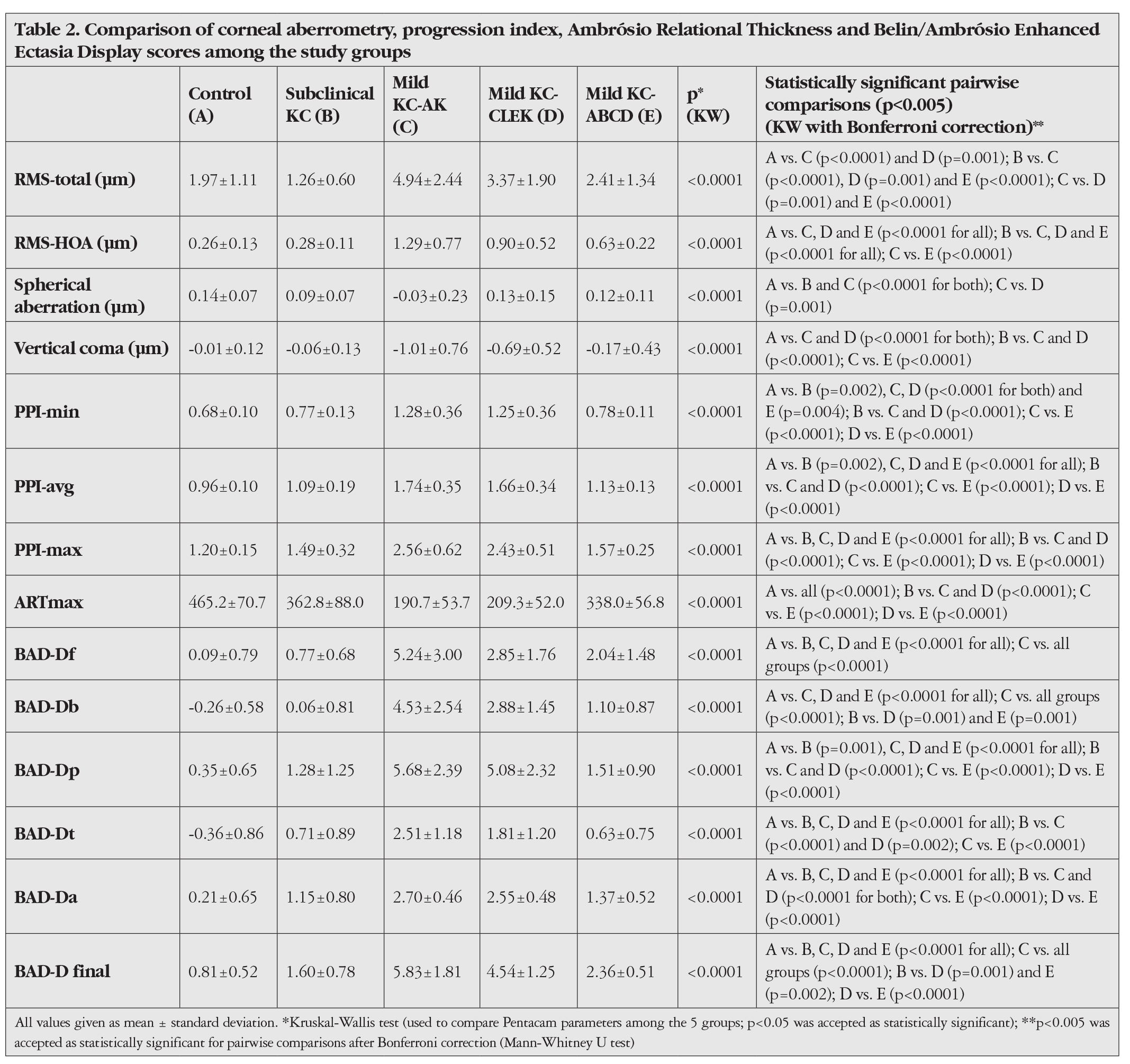
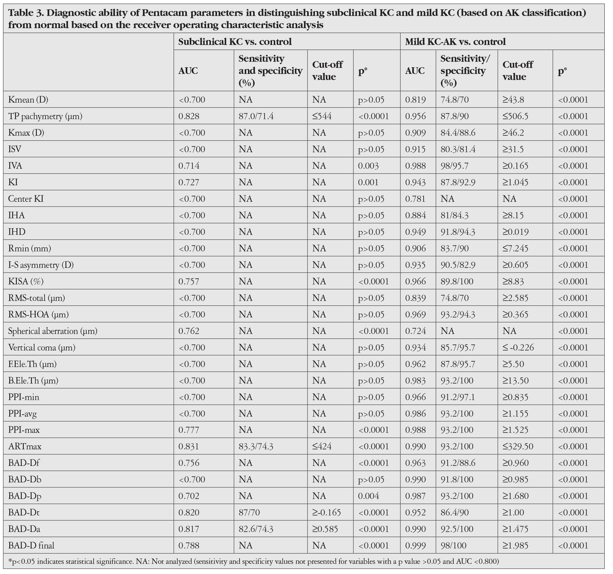
There was no statistically significant difference in age among the control (n=70 eyes; 27.1±9.9 years), subclinical KC (n=24 eyes; 26.2±6.1 years), mild KC-AK (n=101 eyes; 30.2±9.5 years), mild KC-CLEK (n=28 eyes; 29.4±14.4 years), and mild KC-ABCD (n=15 eyes; 29.3±7.5 years) groups (p=0.060, Kruskal-Wallis test). However, Kmean, TP pachymetry, Kmax, ISV, IVA, KI, CKI, IHA, IHD, Rmin, I-S, KISA, RMS values (total, HOA, spherical, and vertical coma aberrations), F.Ele.Th, B.Ele.Th, PPI (min, avg, and max), ARTmax, and BAD-D scores (all) showed significant differences among the five groups (p<0.05, Kruskal-Wallis test, , ).
Discussion
The current study provides a comprehensive re-evaluation of Pentacam parameters in the diagnosis of mild and subclinical KC, also comparing with the earlier publications that utilized a variety of patient selection criteria and definitions. This study demonstrated that the efficacy of Pentacam parameters in diagnosing mild KC was influenced by differences in the “mild KC” criteria between the AK and CLEK classification systems. The present study also demonstrated the performance of Pentacam metrics in identifying eyes with KC that were classified as stage 0 by the Belin ABCD progression display.,,
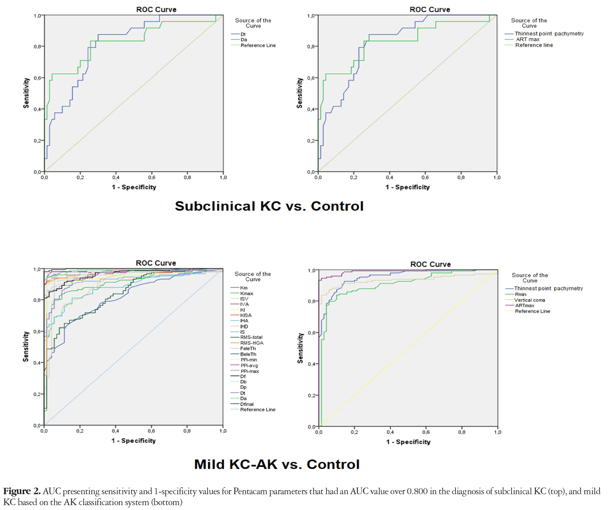
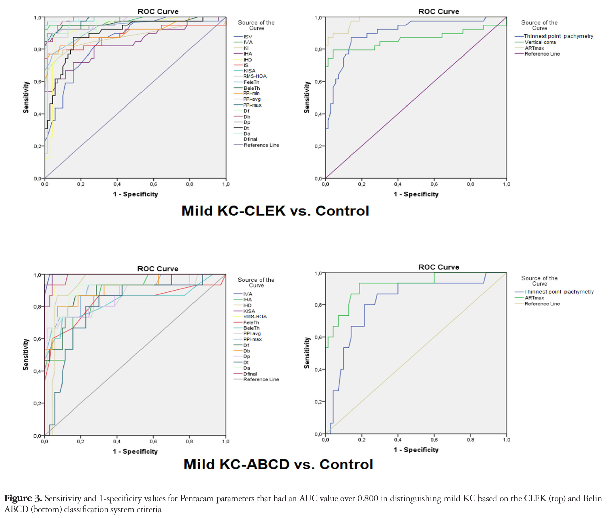
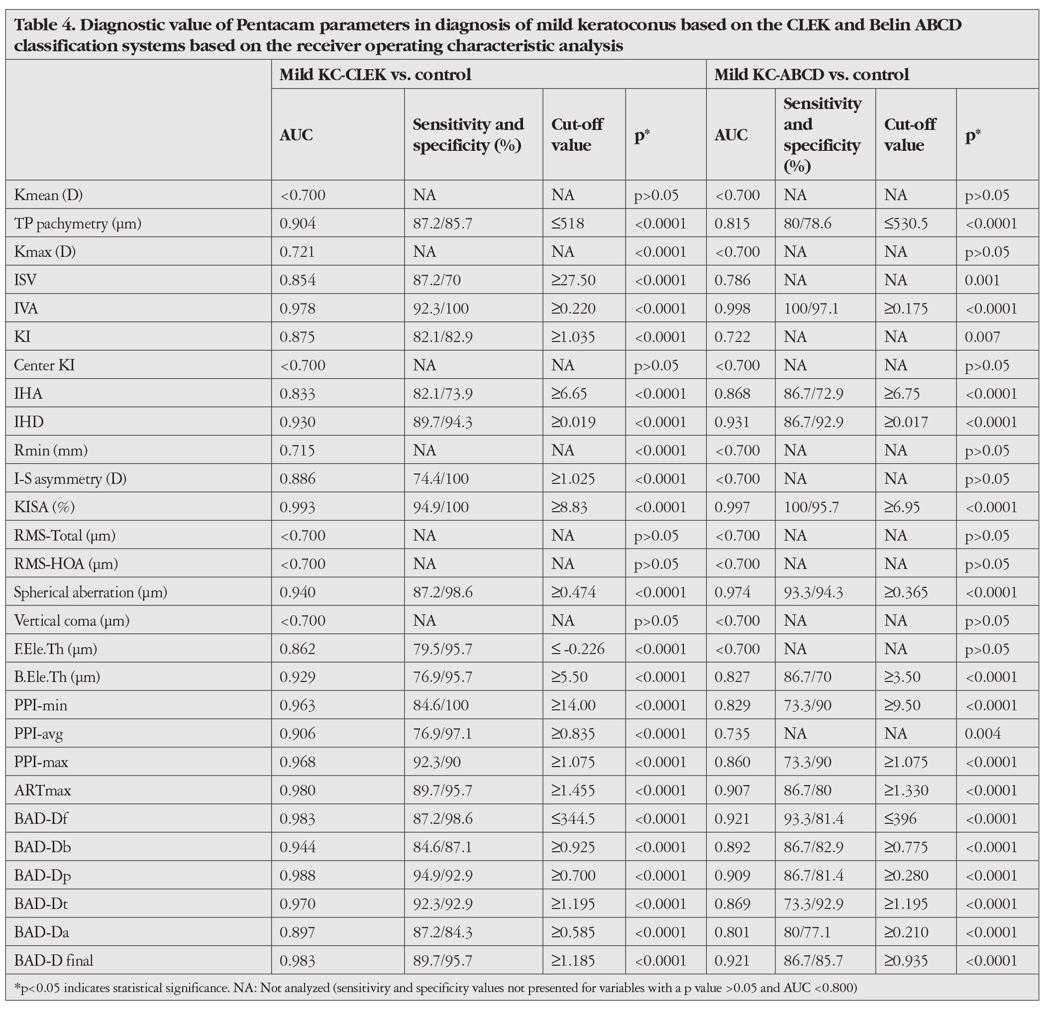
This study showed that among the Pentacam parameters, ARTmax had the highest individual performance in distinguishing eyes with subclinical KC from normal (83.3% sensitivity and 74.3% specificity), followed by TP pachymetry, Dt, and Da. However, D final, KISA, I-S, topometric indices, corneal aberrations, and elevation values had no or poor utility in the detection of subclinical KC. In contrast, most of the Pentacam parameters showed highly satisfying performance in the diagnosis of mild KC, although the most powerful Pentacam parameters and their sensitivity/specificity differed depending on the definition of “mild KC” used. For instance, final D score showed excellent performance in the detection of mild KC based on both the AK (98% sensitivity and 100% specificity) and CLEK (97.4% sensitivity and 100% specificity) definitions when the threshold value was ≥1.985. However, when the ABCD stage 0 descriptors were used, IVA accurately detected all eyes (100%) with KC and 97.1% of the normal eyes, for which D final had 93.3% sensitivity and 95.7% specificity. It should also be noted that ARTmax, KISA, IVA, IHD, RMS-HOA, Da, Db, and PPI-max were the common (for all mild KC-AK, -CLEK and -ABCD groups) powerful Pentacam parameters that showed very high performance (AUC>0.900) in the diagnosis of mild KC.
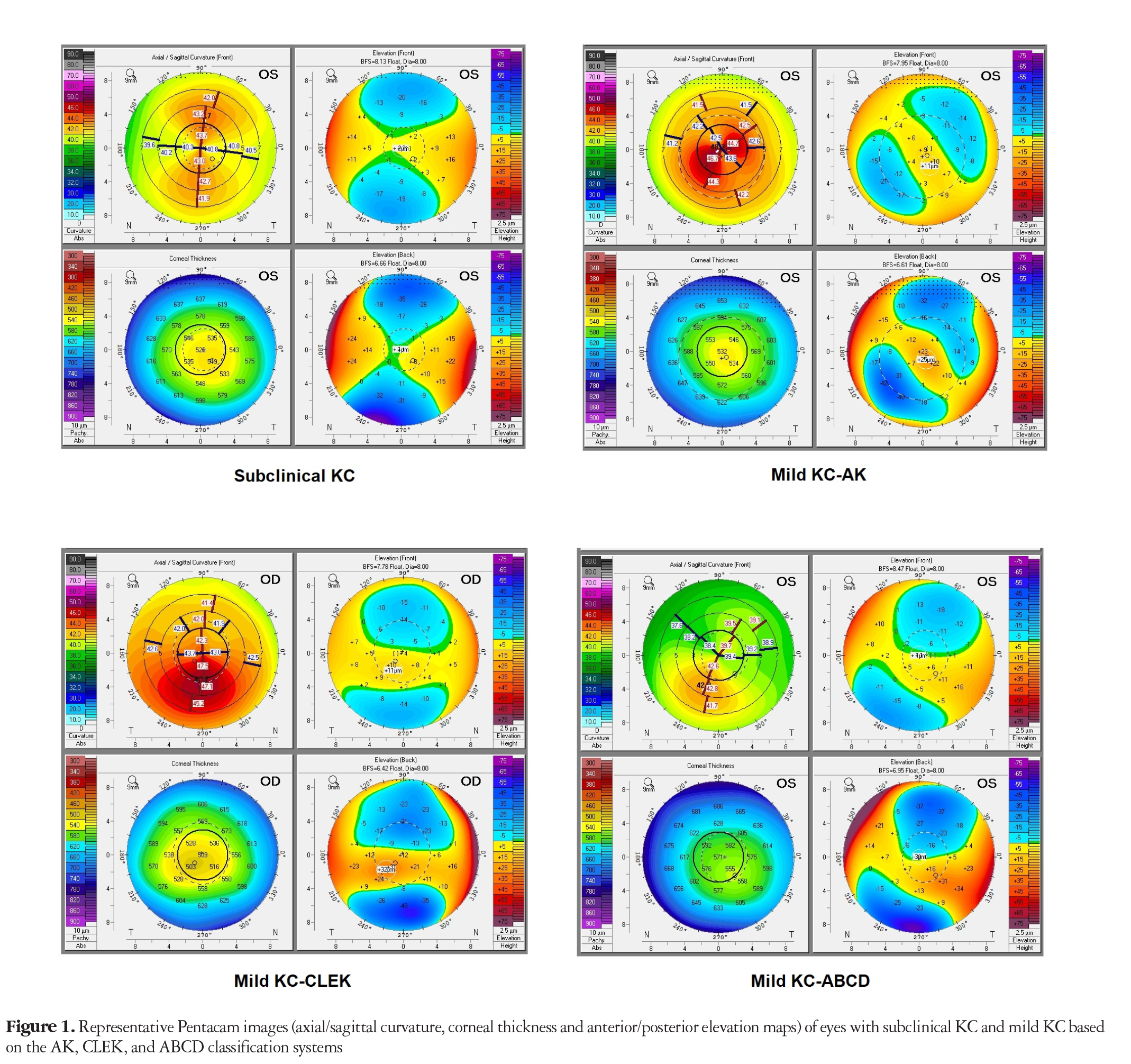
In agreement with the most common definitions of subclinical KC, all eyes in our subclinical KC group had 20/20 corrected vision and normal biomicroscopy, keratometry, and pachymetry but subtle tomographical alterations not reaching the threshold for KC diagnosis.,, Therefore, the subclinical KC group in the current study was able to represent real-world risky cases for laser refractive surgery.
In the present study, ARTmax, TP pachymetry, Dt, and Da had the best sensitivity and specificity values (range: 82.6%-87% and 70%-74.3%, respectively) in subclinical KC detection. Interestingly, these parameters were all associated with corneal thickness and its distribution, suggesting that corneal thickness-related Pentacam data might be particularly useful in the diagnosis of subclinical KC. The sensitivity and specificity values for the Pentacam parameters found in this study were similar to those reported in published studies on subclinical KC diagnosis, which were summarized as follows: 82%-90.5% and 70%-86.5% for ARTmax, 89.2% and 90.3% for Da, and 52.6%-95.5% and 32.4%-94.1% for D final (Supplementary Table S1).,,,,,,,,,,, On the other hand, it can be seen in Supplementary Table S1 that there were overlaps among the criteria for “subclinical KC” and “mild KC”. For instance, Heidari et al. included clinically normal eyes with anterior elevation >12 µm, posterior elevation >17 µm, SRAX <20, Kmax >47.2 D (but <48.7 D) and I-S value >1.4 D (but <1.9 D) at the 3-mm radii as subclinical KC, whereas these criteria practically describe mild KC without biomicroscopic findings.
However, it should be pointed out that the present study and related literature review mainly focused on the individual performance of Pentacam parameters in the diagnosis of subclinical and mild KC. Therefore, we do not discuss the topography-based multifactorial regression formulas introduced in previous studies or parameters from other imaging modalities. Nevertheless, it is obvious from the published literature that corneal epithelial imaging, corneal biomechanical measurements (i.e., CorVis ST, Oculus Inc.® and Ocular Response Analyzer, ORA, Reicherts®) and 3-D morphovolumetric analysis have significant value in diagnosing subclinical KC in addition to corneal topography/tomography.,,,,,,,,
In terms of mild KC diagnosis, the majority of studies in the current literature used the AK stage 1 KC criteria, and excellent to good sensitivity/specificity was observed for F.Ele.Th (sensitivity/specificity: 97.8%/94.8%), B.Ele.Th (100%/99.4%), D final (98%-100%/95.9%-100%), IVA (97.8%/95.8%), KI (93.3%/97.9%), and PPI values (Supplementary Table S2).,,,, These values were very similar to those found in the present study in the diagnosis of mild KC based on the AK, CLEK, and ABCD criteria (sensitivity/specificity ranged from 87.2% to 97.4%/92.9% to 100%). These results might indicate that Pentacam parameters are able to detect an eye with mild KC with high sensitivity regardless of the presence of biomicroscopic signs.
Regarding the “mild” KC-ABCD group in the current study, the ABCD system was actually developed by Belin and Duncan to track KC progression, and stage 0 theoretically describes “normal” eyes. However, we detected 15 eyes with mild KC (all had typical topographical map patterns for KC and the contralateral eye had manifest KC) that were labelled as “stage 0” by the Pentacam ABCD system in our database. These eyes were also included in the present study as the “mildest” KC stage for the ABCD grading system instead of using cases with ABCD stage 1 KC, since Belin and Duncan already reported that ABCD stages 1-4 were closely matched with the AK stages 1-4 in terms of anterior curvature. However, assuming “stage 0” as the mildest grade in the ABCD system may have led to the selection of milder KC cases compared to the mild KC-AK and -CLEK groups. Therefore, the diagnostic performance of the Pentacam parameters might have been underestimated in the mild KC-ABCD group. On the other hand, although the size of the mild KC-ABCD group was relatively small due to its rarity, to our knowledge there is no other study testing the diagnostic performance of Pentacam parameters in the detection of keratoconic eyes categorized as “ABCD stage 0.” One exception is a study by Zhang et al., who used the ABCD stage 0 criteria as “topographic normality” for their forme fruste KC group, which could have led to the inaccurate classification of keratoconic eyes as normal.
Conclusion
As a result of the non-uniform definitions and selection criteria employed in the literature, sensitivity and specificity values show substantial variation in the diagnosis of “subclinical KC.” The current study revealed that corneal thickness-related Pentacam parameters might have value for detecting subclinical KC. However, even with this sophistication, Pentacam has modest capability in the diagnosis of subclinical KC, and further approaches such as corneal biomechanical assessment, epithelial mapping, and 3-D morphovolumetric analysis, which provide robust data on subclinical alterations in the cornea, appear to be necessary.,,,,,,,,
On the other hand, this study also confirmed that Pentacam is able to detect eyes with mild KC with high accuracy, despite the fact that the most powerful parameters have varying specificities and sensitivities depending on the “mild KC” criteria used. Nevertheless, definitive and objective criteria for grading subclinical and clinical KC are essential to attain a global consensus regarding the early diagnosis and management of KC, and clinicians should follow a multi-diagnostic strategy rather than relying solely on Pentacam data prior to corneal refractive surgery.
Study Limitations
The relatively small number of cases in the subclinical KC group might be considered a limitation of the current study. This study also did not include eyes with “forme fruste KC,” which in the majority of the existing literature describes “a clinically and topographically normal eye with manifest KC in the contralateral eye.” The term “forme fruste” was first proposed by Amsler to define unilateral cases as an incomplete, abortive, or atypical form of KC. This conclusion was made mostly due to the fact that unilateral KC is genetically described as a form of autosomal dominant transmission with complete penetrance but partial expression, and if individuals are followed for long enough, the opposite eye may eventually show evidence of KC. In 2015, the Global Consensus on Keratoconus and Ectatic Diseases agreed that environmental, biomechanical, genetic, and biochemical anomalies all contribute to the pathogenesis of KC and true unilateral KC does not exist. However, a recent report by Saad et al. presented a case of stable “unilateral KC” with the longest follow-up period of 14 years.
Abbreviations
ABCD: Belin ABCD classification system, AK: Amsler-Krumeich classification, ARC: Anterior average radii of curvature, ART: Ambrósio Relational Thickness, AUC: Area under the receiver operating characteristic curve, BAD-D: Belin/Ambrósio Enhanced Ectasia Display scores (Df, Db, Dp, Dt, Da, and D final), B.Ele.Diff: Back elevation difference, B.Ele.Th: Back elevation at the thinnest point, CDVA: Corrected distance visual acuity, CLEK: Collaborative Longitudinal Evaluation of Keratoconus study, D: Diopters, F.Ele.Diff: Front elevation difference, F.Ele.Th: Front elevation at the thinnest point, HOA: Higher-order aberrations, IHA: Index of height asymmetry, IHD: Index of height decentration, I-S: Inferior-superior keratometric difference at 3-mm radii, ISV: Index of surface variance, IVA: Index of vertical asymmetry, K: Keratometry, KC: Keratoconus, KI: Keratoconus index, KISA: KC percentage index, LogMAR: Logarithm of the minimal angle of resolution, PPI: pachymetric progression index, PRC: Posterior average radii of curvature, Rmin: Minimum radius of curvature, RMS: Root mean square, SRAX: Skewed radial axes, ST-IN: Superotemporal-inferonasal asymmetry, TKC: Topographical Keratoconus Classification, TP: Thinnest point, Kmax: Maximum keratometry, SD: Standard deviation
References
Zhang X, Munir SZ, Sami Karim SA, Munir WM. A review of imaging modalities for detecting early keratoconus. Eye (Lond). 2021;35:173-187.
Thulasidas M, Teotia P. Evaluation of corneal topography and tomography in fellow eyes of unilateral keratoconus patients for early detection of subclinical keratoconus. Indian J Ophthalmol. 2020;68:2415-2420.
Heidari Z, Mohammadpour M, Hashemi H, Jafarzadehpur E, Moghaddasi A, Yaseri M, Fotouhi A. Early diagnosis of subclinical keratoconus by wavefront parameters using Scheimpflug, Placido and Hartmann-Shack based devices. Int Ophthalmol. 2020;40:1659-1671.
Toprak I, Vega A, Alió Del Barrio JL, Espla E, Cavas F, Alió JL. Diagnostic Value of Corneal Epithelial and Stromal Thickness Distribution Profiles in Forme Fruste Keratoconus and Subclinical Keratoconus. Cornea. 2021;40:61-72.
Toprak I, Cavas F, Velázquez JS, Alio Del Barrio JL, Alio JL. Subclinical keratoconus detection with three-dimensional (3-D) morphogeometric and volumetric analysis. Acta Ophthalmol. 2020;98:933-942.
Ambrósio R Jr, Lopes BT, Faria-Correia F, Salomão MQ, Bühren J, Roberts CJ, Elsheikh A, Vinciguerra R, Vinciguerra P. Integration of Scheimpflug-Based Corneal Tomography and Biomechanical Assessments for Enhancing Ectasia Detection. J Refract Surg. 2017;33:434-443.
Gomes JA, Tan D, Rapuano CJ, Belin MW, Ambrósio R Jr, Guell JL, Malecaze F, Nishida K, Sangwan VS; Group of Panelists for the Global Delphi Panel of Keratoconus and Ectatic Diseases. Global consensus on keratoconus and ectatic diseases. Cornea. 2015;34:359-369.
Henriquez MA, Hadid M, Izquierdo L Jr. A Systematic Review of Subclinical Keratoconus and Forme Fruste Keratoconus. J Refract Surg. 2020;36:270-279.
Amsler M. Die “forme fruste” des Keratokonus [The “forme fruste” of keratoconus]. Wien Klin Wochenschr. 1961;73:842-843
Shetty R, Rao H, Khamar P, Sainani K, Vunnava K, Jayadev C, Kaweri L. Keratoconus Screening Indices and Their Diagnostic Ability to Distinguish Normal From Ectatic Corneas. Am J Ophthalmol. 2017;181:140-148.
Hashemi H, Beiranvand A, Yekta A, Maleki A, Yazdani N, Khabazkhoob M. Pentacam top indices for diagnosing subclinical and definite keratoconus. J Curr Ophthalmol. 2016;28:21-26.
Muftuoglu O, Ayar O, Hurmeric V, Orucoglu F, Kılıc I. Comparison of multimetric D index with keratometric, pachymetric, and posterior elevation parameters in diagnosing subclinical keratoconus in fellow eyes of asymmetric keratoconus patients. J Cataract Refract Surg. 2015;41:557-565.
Uçakhan ÖÖ, Cetinkor V, Özkan M, Kanpolat A. Evaluation of Scheimpflug imaging parameters in subclinical keratoconus, keratoconus, and normal eyes. J Cataract Refract Surg. 2011;37:1116-1124.
Kamiya K, Ishii R, Shimizu K, Igarashi A. Evaluation of corneal elevation, pachymetry and keratometry in keratoconic eyes with respect to the stage of Amsler-Krumeich classification. Br J Ophthalmol. 2014;98:459-463.
McMahon TT, Szczotka-Flynn L, Barr JT, Anderson RJ, Slaughter ME, Lass JH, Iyengar SK; CLEK Study Group. A new method for grading the severity of keratoconus: the Keratoconus Severity Score (KSS). Cornea. 2006;25:794-800.
Alió JL, Piñero DP, Alesón A, Teus MA, Barraquer RI, Murta J, Maldonado MJ, Castro de Luna G, Gutiérrez R, Villa C, Uceda-Montanes A. Keratoconus-integrated characterization considering anterior corneal aberrations, internal astigmatism, and corneal biomechanics. J Cataract Refract Surg. 2011;37:552-568.
Zadnik K, Barr JT, Edrington TB, Everett DF, Jameson M, McMahon TT, Shin JA, Sterling JL, Wagner H, Gordon MO. Baseline findings in the Collaborative Longitudinal Evaluation of Keratoconus (CLEK) Study. Invest Ophthalmol Vis Sci. 1998;39:2537-2546.
Belin MW, Duncan JK. Keratoconus: The ABCD Grading System. Klin Monbl Augenheilkd. 2016;233:701-707.
Bae GH, Kim JR, Kim CH, Lim DH, Chung ES, Chung TY. Corneal topographic and tomographic analysis of fellow eyes in unilateral keratoconus patients using Pentacam. Am J Ophthalmol. 2014;157:103-109.
Steinberg J, Aubke-Schultz S, Frings A, Hülle J, Druchkiv V, Richard G, Katz T, Linke SJ. Correlation of the KISA% index and Scheimpflug tomography in ‘normal’, ‘subclinical’, ‘keratoconus-suspect’ and ‘clinically manifest’ keratoconus eyes. Acta Ophthalmol. 2015;93:199-207.
Ruiseñor Vázquez PR, Galletti JD, Minguez N, Delrivo M, Fuentes Bonthoux F, Pförtner T, Galletti JG. Pentacam Scheimpflug tomography findings in topographically normal patients and subclinical keratoconus cases. Am J Ophthalmol. 2014;158:32-40.
Huseynli S, Abdulaliyeva F. Evaluation of Scheimpflug Tomography Parameters in Subclinical Keratoconus, Clinical Keratoconus and Normal Caucasian Eyes. Turk J Ophthalmol. 2018;48:99-108.
Atalay E, Özalp O, Erol MA, Bilgin M, Yıldırım N. A Combined Biomechanical and Tomographic Model for Identifying Cases of Subclinical Keratoconus. Cornea. 2020;39:461-467.
Koc M, Tekin K, Kiziltoprak H, Inanc M, Kosekahya P, Ozulken K, Durukan I. Topometric and Tomographic Evaluation of Subclinical Keratoconus. Ophthalmic Epidemiol. 2020;27:289-297.
Cui J, Zhang X, Hu Q, Zhou WY, Yang F. Evaluation of Corneal Thickness and Volume Parameters of Subclinical Keratoconus Using a Pentacam Scheimflug System. Curr Eye Res. 2016;41:923-926.
Reinstein DZ, Gobbe M, Archer TJ, Silverman RH, Coleman DJ. Epithelial, stromal, and total corneal thickness in keratoconus: three-dimensional display with artemis very-high frequency digital ultrasound. J Refract Surg. 2009;25:604-610.
Tian L, Zhang D, Guo L, Qin X, Zhang H, Zhang H, Jie Y, Li L. Comparisons of corneal biomechanical and tomographic parameters among thin normal cornea, forme fruste keratoconus, and mild keratoconus. Eye Vis (Lond). 2021;8:44.
Zhang M, Zhang F, Li Y, Song Y, Wang Z. Early Diagnosis of Keratoconus in Chinese Myopic Eyes by Combining Corvis ST with Pentacam. Curr Eye Res. 2020;45:118-123.
Kataria P, Padmanabhan P, Gopalakrishnan A, Padmanaban V, Mahadik S, Ambrósio R Jr. Accuracy of Scheimpflug-derived corneal biomechanical and tomographic indices for detecting subclinical and mild keratectasia in a South Asian population. J Cataract Refract Surg. 2019;45:328-336.
Augustin VA, Son HS, Baur I, Zhao L, Auffarth GU, Khoramnia R. Detecting subclinical keratoconus by biomechanical analysis in tomographically regular keratoconus fellow eyes. Eur J Ophthalmol. 2021:11206721211063740.
Lim HB, Tan GS, Lim L, Htoon HM. Comparison of keratometric and pachymetric parameters with Scheimpflug imaging in normal and keratoconic Asian eyes. Clin Ophthalmol. 2014;8:2215-2220.
Saad A, Rizk M, Gatinel D. Fourteen years follow-up of a stable unilateral Keratoconus: unique case report of clinical, tomographical and biomechanical stability. BMC Ophthalmol. 2022;22:245.

