

Abstract
In this study, we aimed to present a rare case of pulsatile proptosis due to sphenoid wing dysplasia without the features of neurofibromatosis type 1 (NF1). A 17-year-old male patient presented with swelling in the superotemporal region of the right eye. Physical examination revealed facial asymmetry with a pulsatile, ill-defined, soft lesion with in the superotemporal region of the right orbit associated with pulsatile proptosis, downward dystopia, and hypotropia. Computer tomography imaging to establish a differential diagnosis showed temporal lobe herniation secondary to sphenoid wing dysplasia. The patient was assessed for NF1, which is most commonly associated with sphenoid wing dysplasia, but no evidence supporting the diagnosis was found. Patients presenting with proptosis should be carefully examined for pulsation and murmurs, and a trauma history should be investigated. Radiological imaging should be used to facilitate the differential diagnosis, and the current clinical condition should be managed with a multidisciplinary approach.
Keywords: Dystopia, exophthalmos, neurofibromatosis type 1, pulsatile proptosis, sphenoid wing dysplasia
Introduction
Proptosis is defined as one or both eyes bulging forward. It can result from different conditions such as infectious, inflammatory, vascular, and neoplastic conditions. Differential diagnosis is critical since it may risk vision and even life.
Encephalocele is one of the causes of proptosis. Encephaloceles are acquired or congenital herniations of meninges and brain parenchyma through a structural cranial bone defect. Traumatic intraorbital encephalocele, though rare, is the most common type and arises from orbital roof fractures caused by blunt trauma. Congenital cases are often linked with sphenoid dysplasia, which is most frequently associated with neurofibromatosis type 1 (NF1)., Sphenoid dysplasia-related encephaloceles cause ocular symptoms such as dystopia, strabismus, and optic disc pressure. NF1 is a hereditary neurocutaneous disorder. Instances of sphenoid dysplasia occurring without associated NF1 features are rare.
The aim of this article is to present a rare case of non-NF1-related pulsatile proptosis and review of literature.
Case Report
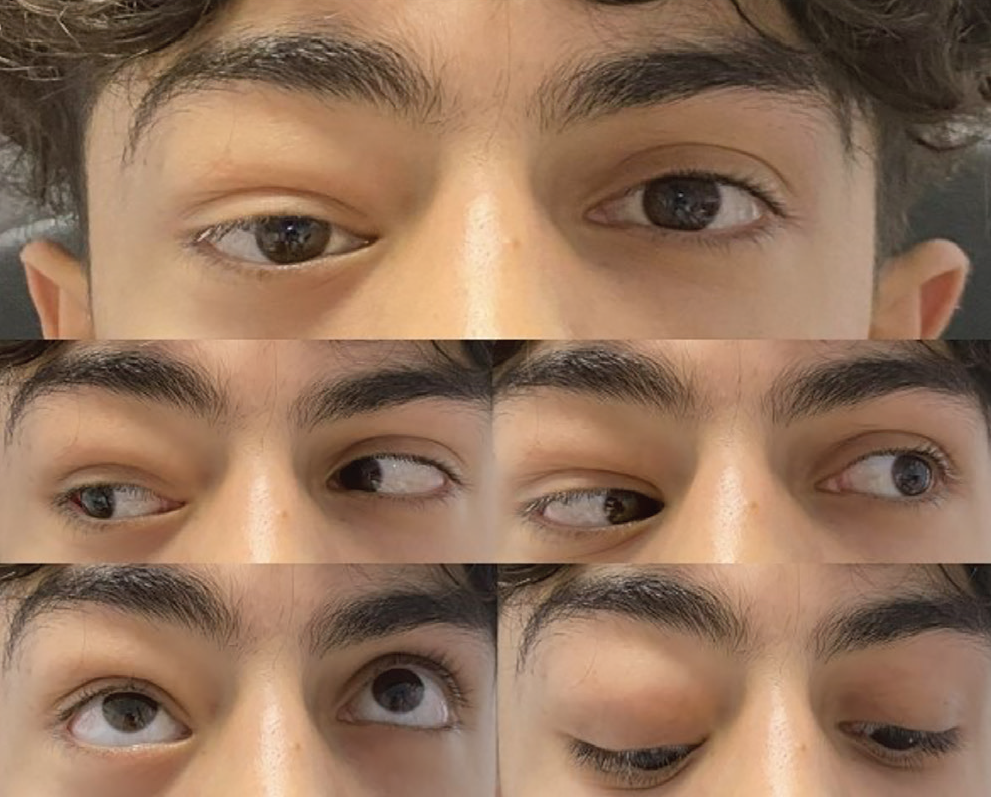
A 17-year-old male patient presented to the eye clinic with a pulsating lesion in the superotemporal region of the right orbit persisting for 5 years. Macroscopic examination revealed facial asymmetry with a pulsatile, ill-defined, soft lesion in the superotemporal region of the right orbit, and no audible bruit was detected.
The patient exhibited downward dystopia, 10 prism diopters of hypotropia in primary position, and proptosis of the right eye (). Eye movements were restricted upward in the right eye but free in other directions. Suppression was observed in the Worth 4-dot test and Schober’s test.
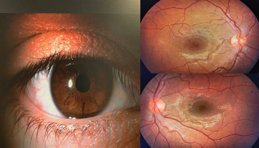
On examination, the patient’s direct and consensual light reflexes were normal, bilateral visual acuity was 20/20 and intraocular pressures were within normal limits. Anterior segment and fundus examination findings were normal (). Optical coherence tomography, retinal nerve fiber layer analysis, and visual field examination revealed no pathology.
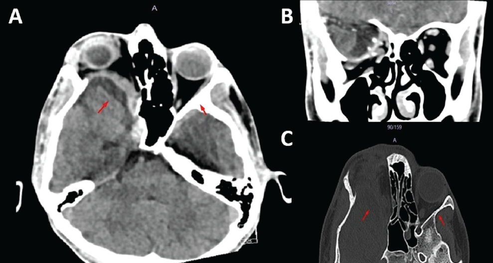
In contrast-free orbital computer tomography, the right greater wing of the sphenoid bone appeared dysplastic (). There was a distinct extra-axial cerebrospinal fluid space in the right temporal fossa, and herniation of the temporal lobe into the orbit markedly displaced the superior oblique muscle. The patient was evaluated for NF1, but there were no findings that satisfied the diagnostic criteria other than sphenoid wing dysplasia. The patient also had no family history of NF1. The family was referred for genetic counseling and no specific feature was identified.
Consultations with the plastic and reconstructive surgery and neurosurgery departments were sought for the patient. Orbital roof reconstruction was recommended by the neurosurgery team. However, the patient declined surgical treatment. He is currently continuing follow-up in the strabismus unit.
Discussion
In this report, we present a rare instance in which a patient presented with pulsatile proptosis and diagnosed with sphenoid wing dysplasia despite having no clinical evidence of NF1.
Proptosis refers to the forward protrusion of one or both eyes from the orbit. This condition arises due to an increase in the contents within the fixed anatomical structure of the bony orbit. The reflection of cerebrospinal fluid pulsations into the orbit due to a defect in the orbital roof is termed pulsatile proptosis. Pulsatile proptosis is observed in conditions such as carotid-cavernous fistula, arteriovenous malformation, and encephalocele secondary to sphenoid wing dysplasia or orbital roof fracture., A differential diagnosis can be made through physical examination, anamnesis (including trauma history, chemosis, epibulbar venous congestion, and the absence of a bruit), and radiological imaging. Our patient had pulsatile proptosis but no trauma history, chemosis, epibulbar venous congestion, or bruit.
Sphenoid wing dysplasia is characterized by hypoplasia or total absence of the greater or lesser wing of the sphenoid bone. It leads to the expansion of the orbit and middle cranial fossa, causing herniation of the temporal lobe into the orbit. Pulsatile proptosis is observed as a result. Although it can be seen as an isolated deformity, it most commonly occurs in association with NF1.
Sphenoid wing dysplasia is one of the major diagnostic criteria for NF1, and is considered a pathognomonic finding for the disease. Between 5% and 12% of NF1 patients have signs of sphenoid wing dysplasia. NF1 is an autosomal dominant inherited disorder that arises from a mutation of a gene of chromosome 17q11.2. NF1 diagnostic criteria include: >6 cafe-au-lait macules, >2 neurofibromas, freckling in axillary or inguinal regions, optic pathway glioma, >2 iris Lisch nodules or choroid abnormalities, distinctive osseous lesion (such as sphenoid dysplasia, pseudoarthrosis of a long bone), presence of a heterozygous pathogenic NF1 variant. NF1 is characterized by the presence of two or more of the diagnostic criteria. In our patient, there were no findings that satisfied the diagnostic criteria other than sphenoid wing dysplasia, and there was no family history of NF1.
Upon reviewing the literature, we found that only 5 patients with sphenoid wing dysplasia did not have evidence of NF1. Among these, one patient was diagnosed with myofibroma, another with craniofacial fibrous dysplasia, and the other three patients had isolated bone defects, similar to our case., , summarizes case reports of sphenoid wing dysplasia in the literature., ,,,,,,,,,,,,,,,,,,,,,,
Table 1. Case reports of sphenoid wing dysplasia with pulsatile proptosis in the literature are summarized by article title, patient sex and age, and etiology
Age (years) | Sex | Etiology | Articleref# |
|---|---|---|---|
7 | M | Isolated bone defect | Sphenoid wing dysplasia: report of 3 cases10 |
36 | M | NF1 | |
19 | M | Craniofacial fibrous dysplasia | |
43 | M | NF1 | Traumatic brain injury, bulging eyeball, and skin lumps13 |
19 | F | NF1 | Acute enophthalmos after lumbar puncture in a patient with type 1 neurofibromatosis related sphenoid wing dysplasia14 |
14 | M | NF1 | Symptomatic enophthalmos due to sphenoid wing dysplasia appearing over 12 years in a patient with neurofibromatosis type 1: a case report and literature review15 |
Unknown | Unknown | NF1 (4 cases) | Reconstruction of sphenoid wing dysplasia in neurofibromatosis type 1 patients: an evolving technique16 |
41 | F | NF1 | Pulsating proptosis and heavy eye syndrome precipitated by neurofibromatosis type 1: a case report17 |
25 | F | NF1 | Computer-aided three-dimensional virtual surgical planning in complex skull base reconstruction for sphenoid wing dysplasia in neurofibromatosis type 118 |
18 | M | Isolated bone defect | Sphenoid wing dysplasia in the absence of neurofibromatosis: Diagnosis and management of a novel phenotype4 |
20 | M | NF1 | Neuroimaging findings of extensive sphenoethmoidal dysplasia in NF119 |
59 | F | NF1 | Imaging findings of jugular foramen meningocele in a neurofibromatosis type 1 patient20 |
1 week | M | Myofibroma | Sphenoid dysplasia: a rare presentation of infantile myofibroma21 |
38 | F | NF1 | A multidisciplinary approach to sphenoid wing dysplasia presenting with pulsatile proptosis in neurofibromatosis type 1: a rare case report22 |
9 | M | NF1 | Evidence of neurofibromatosis type 1 in a multi-morbid Inca child mummy: a paleoradiological investigation using computed tomography23 |
2 | M | NF1 | Sphenoid wing dysplasia with pulsatile exophthalmos in neurofibromatosis type 124 |
13 | F | NF1 | Sphenoid wing dysplasia and plexiform neurofibroma in neurofibromatosis type 125 |
57 | F | NF1 | Ipsilateral sphenoid wing dysplasia, orbital plexiform neurofibroma and fronto-parietal dermal cylindroma in a patient with segmental neurofibromatosis26 |
1 month | M | NF1 | A rare case of primary congenital glaucoma in combination with neurofibromatosis 1: a case report27 |
55 | F | NF1 | Increased ocular pulse amplitude associated with unilateral dysgenesis of the orbital roof11 |
55 | F | Isolated bone defect | |
15 | M | NF1 | Treatment of sphenoid dysplasia with a titanium-reinforced porous polyethylene implant in orbitofrontal neurofibroma: report of three cases28 |
18 | M | NF1 | |
25 | M | NF1 | |
6 | M | NF1 | Orbital reconstruction for pulsatile exophthalmos secondary to sphenoid wing dysplasia12 |
7 | M | NF1 | Reconstruction of skull base defects in sphenoid wing dysplasia associated with neurofibromatosis 1 with titanium mesh29 |
21 | F | NF1 | |
30 | F | NF1 | Reconstruction of the sphenoid wing in a case of neurofibromatosis type 1 and complex unilateral orbital dysplasia with pulsating exophthalmos30 |
25 | M | NF1 | Reconstruction of sphenoid wing dysplasia with pulsating exophthalmos in a case of neurofibromatosis type 1 supported by intraoperative navigation using a new skull reference system31 |
15 months | F | NF1 | Cranio-orbital-temporal neurofibromatosis: are we treating the whole problem?32 |
M: Male, F: Female, NF1: Neurofibromatosis type 1
In the literature, there are publications reporting orbital roof reconstruction with a titanium mesh and/or bone graft in the treatment, although they are limited in number. Surgeries performed early after diagnosis have been shown to have a more successful visual prognosis.,
In conclusion, history of trauma should not be overlooked in the differential diagnosis of pulsatile lesions around the orbit, and radiological imaging should be utilized to aid diagnosis. Additionally, findings related to NF1, which are closely linked to the patient’s symptoms, should be thoroughly examined, and genetic counseling should be provided to the patient.
Footnotes
Ethics
Informed Consent: Obtained.
Authorship Contributions
Surgical and Medical Practices: S.S.İ, M.E., Concept: S.S.İ, M.E., Design: M.A., H.M., Data Collection or Processing: Y.D.A., Analysis or Interpretation: M.A., H.M., Literature Search: Y.D.A., M.E., Writing: Y.D.A., M.E.
Conflict of Interest
Conflict of Interest: No conflict of interest was declared by the authors.
Financial Disclosure
Financial Disclosure: The authors declared that this study received no financial support.
References
Teng Siew T, Mohamad SA, Sudarno R, Nilamani V. Unilateral Proptosis and Bilateral Compressive Optic Neuropathy in a Meningioma Patient. Cureus. 2024;16:e53728.
Arslan E, Arslan S, Kalkısım S, Arslan A, Kuzeyli K. Long-Term Results of Orbital Roof Repair with Titanium Mesh in a Case of Traumatic Intraorbital Encephalocele: A Case Report and Review of Literature. Craniomaxillofac Trauma Reconstr. 2016;9:255-259.
Gazioğlu N, Ulu MO, Ozlen F, Uzan M, Ciplak N. Acute traumatic orbital encephalocele related to orbital roof fracture: reconstruction by using porous polyethylene. Ulus Travma Acil Cerrahi Derg. 2008;14:247-252.
Zapatero ZD, Kalmar CL, Kosyk MS, Carlson AR, Bartlett SP. Sphenoid Wing Dysplasia in the Absence of Neurofibromatosis: Diagnosis and Management of a Novel Phenotype. Plast Reconstr Surg Glob Open. 2021;9:e3483.
Butt S, Patel BC. Exophthalmos. 2023 Jun 26. In: StatPearls [Internet]. Treasure Island (FL): StatPearls Publishing; 2024.
Zeitoun IM, Ebeid K, Soliman AY. Growing skull fractures of the orbital roof: a multicentric experience with 28 patients. Childs Nerv Syst. 2021;37:1209-1217.
Ghorbani M, Lafta G, Rahbarian F, Mortazavi A. Treatment of post-traumatic direct carotid-cavernous fistulas using flow diverting stents: Is it alone satisfactory? J Clin Neurosci. 2021;86:230-234.
Antico A, Vitulli F, Rossi A, Gaggero G, Piatelli G, Consales A. Pathogenesis and treatment of a giant occipital bone defect with meningoencephalocele in an NF1 child: case report and review of the literature. Child’s Nerv Syst. 2024;40:27-39.
Legius E, Messiaen L, Wolkenstein P, Pancza P, Avery RA, Berman Y, Blakeley J, Babovic-Vuksanovic D, Cunha KS, Ferner R, Fisher MJ, Friedman JM, Gutmann DH, Kehrer-Sawatzki H, Korf BR, Mautner VF, Peltonen S, Rauen KA, Riccardi V, Schorry E, Stemmer-Rachamimov A, Stevenson DA, Tadini G, Ullrich NJ, Viskochil D, Wimmer K, Yohay K; International Consensus Group on Neurofibromatosis Diagnostic Criteria (I-NF-DC); Huson SM, Evans DG, Plotkin SR. Revised diagnostic criteria for neurofibromatosis type 1 and Legius syndrome: an international consensus recommendation. Genet Med. 2021;23:1506-1513.
Qayyum MU, Haq EU, Mahmood Kazmi SA, Zahra R. Sphenoid Wing Dysplasia: Report Of 3 Cases. Vol. 34(Suppl 1, Journal of Ayub Medical College, Abbottabad: JAMC. Pakistan; 2022. p. S1013-1020.
Vira AS, Mahmoud AM, Roberts CJ, Katz SE. Increased Ocular Pulse Amplitude Associated with Unilateral Dysgenesis of the Orbital Roof. Vol. 6, Case reports in ophthalmology. Switzerland; 2015. p. 158-163.
Dale EL, Strait TA, Sargent LA. Orbital reconstruction for pulsatile exophthalmos secondary to sphenoid wing dysplasia. Ann Plast Surg. 2014;72(6):S107-11.
Yu Q, Zhu J, Wu H. Traumatic Brain Injury, Bulging Eyeball, and Skin Lumps. World Neurosurg. 2022;168:207-208.
Miano DI, Byrd G, Kattoula R, Thet A, Adkins R, Cosgrove R, Johnson SS. Acute Enophthalmos After Lumbar Puncture in a Patient with Type 1 Neurofibromatosis Related Sphenoid Wing Dysplasia. Neuroophthalmology. 2022;46:270-274.
Fujino S, Enokizono M, Tamada I, Ihara S, Kono T, Miyama S. Symptomatic enophthalmos due to sphenoid wing dysplasia appearing over 12 years in a patient with neurofibromatosis type 1: a case report and literature review. J AAPOS. 2022;26:210-214.
Goddard MNV, Dunne MJ, Ghorbanian MS, Eccles MS. Reconstruction of Sphenoid Wing Dysplasia in Neurofibromatosis Type-1 Patients: An Evolving Technique. JPRAS Open. 2022;31:67-71.
Lai YF, Lee LC, Chen YH, Chien KH. “Pulsating proptosis and heavy eye syndrome precipitated by neurofibromatosis type 1: A case report”. Medicine (Baltimore). 2021;100:e27575.
Hsu HH, Wong CE, Lee JW, Huang CC, Lee JS. Computer-Aided Three-Dimensional Virtual Surgical Planning in Complex Skull Base Reconstruction for Sphenoid Wing Dysplasia in Neurofibromatosis Type 1. J Craniofac Surg. 2021;32:2539-2541.
Tam A, Sliepka JM, Bellur S, Bray CD, Lincoln CM, Nagamani SCS. Neuroimaging findings of extensive sphenoethmoidal dysplasia in NF1. Clin Imaging. 2018;51:160-163.
Serindere M, Tasar M, Hamcan S, Bozlar U. Imaging Findings of Jugular Foramen Meningocele in a Neurofibromatosis Type 1 Patient. Case Rep Radiol. 2017;2017:7047696.
Kini A, Syed R, Compton C, Hata JL, Ramasubramanian A. Sphenoid Dysplasia: A Rare Presentation of Infantile Myofibroma. Ophthalmic Plast Reconstr Surg. 2018;34:65-67.
Prathibha S, Parasar V, Yasmin S, Seetha Pramila VV. A multidisciplinary approach to sphenoid wing dysplasia presenting with pulsatile proptosis in neurofibromatosis Type 1: A rare case report. Indian J Ophthalmol. 2018;66:157-160.
Panzer S, Wittig H, Zesch S, Rosendahl W, Blache S, Müller-Gerbl M, Hotz G. Evidence of neurofibromatosis type 1 in a multi-morbid Inca child mummy: A paleoradiological investigation using computed tomography. PLoS One. 2017;12:e0175000.
Rommel FR, Spors H, Grzybowski M, Hahn A, Neubauer BA. Sphenoid Wing Dysplasia with Pulsatile Exophthalmos in Neurofibromatosis Type 1. Neuropediatrics. 2016;47:278-279.
Rennert RC, Scott Pannell J, Levy ML, Khalessi AA. Sphenoid wing dysplasia and plexiform neurofibroma in neurofibromatosis type 1. ANZ J Surg. 2018;88:615-616.
Friedrich RE, Hagel C, Mautner VF. Ipsilateral Sphenoid Wing Dysplasia, Orbital Plexiform Neurofibroma and Fronto-Parietal Dermal Cylindroma in a Patient with Segmental Neurofibromatosis. Anticancer Res. 2015;35:6813-6818.
Li H, Liu T, Chen X, Xie L. A rare case of primary congenital glaucoma in combination with neurofibromatosis 1: a case report. BMC Ophthalmol. 2015;15:149.
Niddam J, Bosc R, Suffee TM, Le Guerinel C, Wolkenstein P, Meningaud JP. Treatment of sphenoid dysplasia with a titanium-reinforced porous polyethylene implant in orbitofrontal neurofibroma: report of three cases. J Craniomaxillofac Surg. 2014;42:1937-1941.
Lotfy M, Xu R, McGirt M, Sakr S, Ayoub B, Bydon A. Reconstruction of skull base defects in sphenoid wing dysplasia associated with neurofibromatosis I with titanium mesh. Clin Neurol Neurosurg. 2010;112:909-914.
Friedrich RE. Reconstruction of the sphenoid wing in a case of neurofibromatosis type 1 and complex unilateral orbital dysplasia with pulsating exophthalmos. In Vivo. 2011;25:287-290.
Friedrich RE, Heiland M, Kehler U, Schmelzle R. Reconstruction of Sphenoid Wing Dysplasia with Pulsating Exophthalmos in a Case of Neurofibromatosis Type 1 Supported by Intraoperative Navigation Using a New Skull Reference System. Skull Base. 2003;13:211-217.
Havlik RJ, Boaz J. Cranio-orbital-temporal neurofibromatosis: are we treating the whole problem? J Craniofac Surg. 1998;9:529-535.

