

Abstract
Objectives
Yasunari nodules are choroidal lesions observed in patients diagnosed with neurofibromatosis type 1 (NF-1) and characterized by relatively irregular dome-shaped, plaque-like, or patchy boundaries. The present study examines the multimodal imaging characteristics of Yasunari nodules and their value in the diagnosis of NF-1.
Materials and Methods
Medical records including optical coherence tomography (OCT), enhanced depth imaging OCT, infrared reflectance (IR) imaging, OCT angiography, and color fundus images of NF-1 patients who were examined at the Department of Ophthalmology in Dokuz Eylül University Faculty of Medicine between January 2022 and December 2023 were retrospectively reviewed for the presence of Yasunari nodules.
Results
A total of 54 eyes of 27 patients were included in the study. At least one choroidal nodule was detected on IR imaging in 52 eyes (96.3%). In 31 (72.1%) of the 43 eyes (79.6%) with available high-quality OCT angiography images, choroidal nodules were observed as areas showing a flow deficit in the choriocapillaris layer. Of the total 54 eyes included, Lisch nodules without choroidal nodules were observed in 2 eyes (3.7%). In 16 eyes (29.6%), Lisch nodules were not detected despite the presence of choroidal nodules. Both Lisch nodules and choroidal nodules were detected in the other 36 eyes (66.7%).
Conclusion
Yasunari nodules are frequently observed in NF-1 cases and can be easily detected with multimodal imaging techniques, especially IR imaging. The ability to visualize choroidal nodules before the appearance of Lisch nodules demonstrates the importance of Yasunari nodules in the diagnosis of NF-1.
Keywords: Choroidal nodules, infrared imaging, multimodal imaging, neurofibromatosis type 1, Yasunari nodules
Introduction
Neurofibromatosis type 1 (NF-1) is an autosomal dominant disease caused by deletions or mutations in the neurofibromin gene located on chromosome 17p11.2. It has a reported incidence of approximately 1/3000 and a prevalence between 1/4000 and 1/5000. NF-1 is characterized by a range of findings, including nerve tumors that can develop in various parts of the body, cutaneous pigmentation changes (cafe au lait spots, axillary and inguinal freckling), vascular abnormalities, and bone lesions (pseudoarthrosis, sphenoidal wing hypoplasia), and is also commonly associated with ocular involvement., ,
The NF-1 diagnostic criteria determined by the National Institutes of Health include the presence of two or more Lisch nodules on the iris and optic nerve glioma as ophthalmological findings. In 2021, Legius et al. suggested that the presence of at least two choroidal abnormalities described as “bright, patchy nodules” on optical coherence tomography (OCT) or infrared reflectance (IR) imaging should also be used as a diagnostic criterion for NF-1. These lesions, which occur at the level of the choroid, are hamartomatous nodules also referred to as “Yasunari nodules”. Their reported prevalence in NF-1 cases ranges from 28% to 100%., , ,
The aim of this study was to evaluate the incidence of choroidal nodules in patients with NF-1, their multimodal imaging characteristics, and their place in the diagnosis of NF-1.
Materials and Methods
This retrospective study was conducted within the framework of the principles of the Declaration of Helsinki and approved by the Dokuz Eylül University Local Ethics Committee (approval number: 2023/10-01, date: 29.03.2023). We retrospectively analyzed the medical records and recorded OCT, enhanced depth imaging OCT (EDI-OCT), IR, OCT angiography (OCTA), and color fundus images of patients who were examined in the Dokuz Eylül University Faculty of Medicine, Department of Ophthalmology between January 2022 and December 2023 and had at least two of the National Institutes of Health diagnostic criteria for NF-1. Exclusion criteria were optic media opacity that could interfere with posterior segment imaging, comorbid systemic or ocular disease other than NF-1, and a history of systemic drug use.
All patients included in the study underwent a comprehensive ophthalmological assessment including anterior and posterior segment examination. Spectral-domain OCT images (7-mm fovea-centered radial B-scan sections with a30-degree lens, ART: 9), EDI-OCT images (9-mm horizontal sections passing through the fovea) and IR images (fovea-centered 55-degree area) were obtained with a standard protocol using the Heidelberg Spectralis (Heidelberg Engineering, Heidelberg, Germany) device. OCTA images covering a 12x12-mm area centered on the fovea and/or optic disc were obtained using the DRI OCT Triton (TOPCON, Tokyo, Japan) device, and central 45-degree color fundus images including the optic disc and macula were obtained using the VISUCAM 500 (Carl Zeiss Meditec, Jena, Germany) or DRI OCT Triton device. We recorded the patients’ age, gender, presence of cafe au lait spots, and cranial magnetic resonance imaging (MRI) findings for each patient; central macular thickness (CMT) and presence of Lisch nodules, eyelid plexiform neurofibroma, optic glioma, and Yasunari nodules in each eye; and subfoveal choroidal thickness (SFCT) for eyes with EDI-OCT images passing through the fovea.
Using the device software, CMT was determined by manually measuring the shortest vertical distance between the internal limiting membrane and Bruch’s membrane in OCT images passing through the fovea, and SFCT was determined by manually measuring the shortest vertical distance between the hyperreflective outer border of the retinal pigment epithelium (RPE) and the choroid-sclera junction in EDI-OCT images passing through the fovea.
The presence of optic glioma was evaluated by examining contrast-enhanced orbital MRI.
Lisch nodules were defined as yellowish brown dome-shaped solid hamartomatous lesions 1-2 mm in diameter on the iris surface on slit-lamp examination.
Choroidal nodules were identified on IR images as domed, placoid, or patchy choroidal lesions with relatively irregular borders and high reflectivity. The presence of these nodules was also evaluated on OCT images with image quality ≥18 and OCTA images with image quality ≥40 for patients with recorded images.
Informed consent was not obtained because the study was retrospective and used no identifying data or images.
Statistical Analysis
Statistical analyses were performed using the SPSS statistical program (IBM Corp, Armonk, NY, USA). Descriptive statistics were used to summarize the data. Categorical variables are presented as numbers and percentages, and quantitative variables are presented as mean ± standard deviation.
Results
The study included a total of 54 eyes of 27 patients. Descriptive statistics of the study group are given in ; detailed demographic and clinical characteristics are shown in .
Table 1. Descriptive statistics of the patients and eyes included in the study
Number of patients/eyes, n | 27/54 |
|---|---|
Age, years | 22.33±14.62a |
Gender, M/F | 14/13a |
Cafe au lait spots, n (%) | 27 (100)a |
Lisch nodule, n (%) | 40 (74.1)b |
Eyelid neurofibroma, n (%) | 1 (1.9)b |
Optic glioma, n (%) | 9 (64.3)c |
Central macular thickness, µm | 244.31±25.17b |
Subfoveal choroidal thickness, µm | 320.49±63.63b |
Choroidal nodule on IR imaging, n (%) | 52 (96.3)b |
Choroidal nodule on OCTA, n (%) | 31 (72.1)d |
a: According to number of patients, b: According to number of eyes, c: According to number of patients with contrast-enhanced orbital magnetic resonance imaging, d: According to number of eyes with adequate quality OCTA images, M: Male, F: Female, IR: Infrared reflectance, OCTA: Optical coherence tomography angiography
Table 2. Demographic and clinical characteristics of the patients
Patient | Sex | Age (years) | Cranial MRI findings | Eye | Lisch nodule | Eyelid neurofibroma | Optic glioma | CMT (µm) | SFCT (µm) | Choroidal nodule on IR | Choroidal nodule on OCTA |
|---|---|---|---|---|---|---|---|---|---|---|---|
1 | M | 52 | No cranial MRI | Right Left | + + | - - | - - | 220 380 | 351 354 | + + | + No OCTA |
2 | F | 41 | No pathology | Right Left | + - | - - | - + | 214 209 | 307 299 | + + | + + |
3 | F | 12 | Hamartoma | Right Left | + + | - - | + - | 268 243 | 347 337 | + + | - - |
4 | F | 16 | Hamartoma | Right Left | + + | - - | - - | 242 236 | 387 362 | + + | + + |
5 | F | 16 | Hamartoma | Right Left | + + | - - | + - | 237 228 | 373 408 | + + | + + |
6 | F | 13 | Non-specific hyperintense foci | Right Left | + - | - - | No orbital MRI No orbital MRI | 227 228 | 349 363 | + + | No OCTA No OCTA |
7 | F | 14 | No cranial MRI | Right Left | + + | - - | - - | 232 262 | 345 346 | + + | + + |
8 | M | 10 | Hamartoma | Right Left | - - | - - | No orbital MRI No orbital MRI | 240 237 | 389 384 | + + | + + |
9 | M | 13 | Hamartoma ICA thinning | Right Left | - - | - - | + - | 232 232 | 359 305 | + + | + + |
10 | M | 57 | Encephalomalacia | Right Left | + + | - - | No orbital MRI No orbital MRI | 228 221 | 177 139 | + + | + + |
11 | M | 15 | Venous anomaly | Right Left | + + | - - | No orbital MRI No orbital MRI | 244 250 | 277 356 | + + | + + |
12 | M | 24 | Bone dysplasia Plexiform neurofibroma | Right Left | + + | - + | No orbital MRI No orbital MRI | 249 181 | 352 No EDI-OCT | + + | + No OCTA |
13 | F | 25 | No pathology | Right Left | + + | - - | - - | 237 236 | No EDI-OCT No EDI-OCT | + + | - - |
14 | F | 23 | Hamartoma | Right Left | + + | - - | - - | 282 263 | 323 316 | + + | + + |
15 | M | 48 | Ventricular dilatation | Right Left | + + | - - | No orbital MRI No orbital MRI | 237 238 | 254 224 | + + | + + |
16 | M | 19 | Hamartoma Glial tumor | Right Left | - + | - - | + - | 235 244 | 302 300 | + + | + + |
17 | F | 18 | Hamartoma Chiari type I anomaly | Right Left | + + | - - | No orbital MRI No orbital MRI | 262 255 | No EDI-OCT No EDI-OCT | + + | - - |
18 | F | 11 | Hamartoma Ventricular dilatation | Right Left | + + | - - | + - | 249 256 | 337 313 | + + | + + |
19 | M | 17 | Venous anomaly | Right Left | + + | - - | + - | 248 249 | 330 316 | + + | No OCTA No OCTA |
20 | F | 47 | No cranial MRI | Right Left | - + | - - | No orbital MRI No orbital MRI | 240 250 | 206 167 | + + | No OCTA No OCTA |
21 | M | 19 | Hamartoma | Right Left | - - | - - | No orbital MRI No orbital MRI | 253 268 | 307 271 | + + | + + |
22 | F | 40 | No cranial MRI | Right Left | - - | - - | No orbital MRI No orbital MRI | 251 258 | 284 275 | + + | + + |
23 | F | 12 | Expansile lesion in pons | Right Left | - + | - - | - + | 223 223 | 324 317 | + + | - - |
24 | M | 11 | Non-specific hyperintense foci | Right Left | + + | - - | No orbital MRI No orbital MRI | 244 254 | 359 360 | - - | - - |
25 | M | 10 | No pathology | Right Left | + + | - - | No orbital MRI No orbital MRI | 245 264 | No EDI-OCT No EDI-OCT | + + | No OCTA No OCTA |
26 | M | 12 | Hamartoma Arachnoid cyst | Right Left | + + | - - | + + | 253 248 | 426 469 | + + | - + |
27 | M | 8 | Hamartoma | Right Left | - + | - - | No orbital MRI No orbital MRI | 238 250 | 302 313 | + + | No OCTA No OCTA |
MRI: Magnetic resonance imaging, CMT: Central macular thickness, SFCT: Subfoveal choroidal thickness, IR: Infrared reflectance, OCTA: Optical coherence tomography angiography, ICA: Internal carotid artery, EDI-OCT: Enhanced depth imaging optical coherence tomography
Fourteen (51.9%) of the patients were male and 13 (48.1%) were female. The mean age of the patients was 22.33±14.62 years (range, 8-57 years). All patients (100%) had at least 6 cafe au lait spots.
Lisch nodules were observed on slit-lamp examination in 40 (74.1%) of the 54 eyes. Neurofibroma on the upper eyelid was noted in 1 eye (1.9%) (patient 12, left eye).
Contrast-enhanced cranial MRI was performed in 23 patients (85.2%). Hamartoma was detected in 12 patients (52.2%); non-specific hyperintense foci, venous anomaly, and ventricular dilatation were each detected in 2 patients (8.7%); and thinning of the internal carotid artery, encephalomalacia, sphenoid bone dysplasia, plexiform neurofibroma, glial tumor, Chiari malformation type I, expansile lesion in the pons, and arachnoid cyst were each detected in 1 patient (4.3%). Three patients (12.9%) had no pathological findings.
Of 14 patients (51.8%) who underwent contrast-enhanced orbital MRI, 9 (33.3%) had optic glioma (unilateral in 8 patients [57.1%] and bilateral in 1 patient [7.1%]).
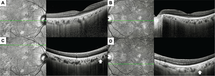
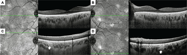
The mean CMT for all eyes was 244.31±25.17 µm (range, 181-380 µm), while the mean SFCT for 47 eyes with EDI-OCT images passing through the fovea was 320.49±63.63 µm (range, 139-469 µm).
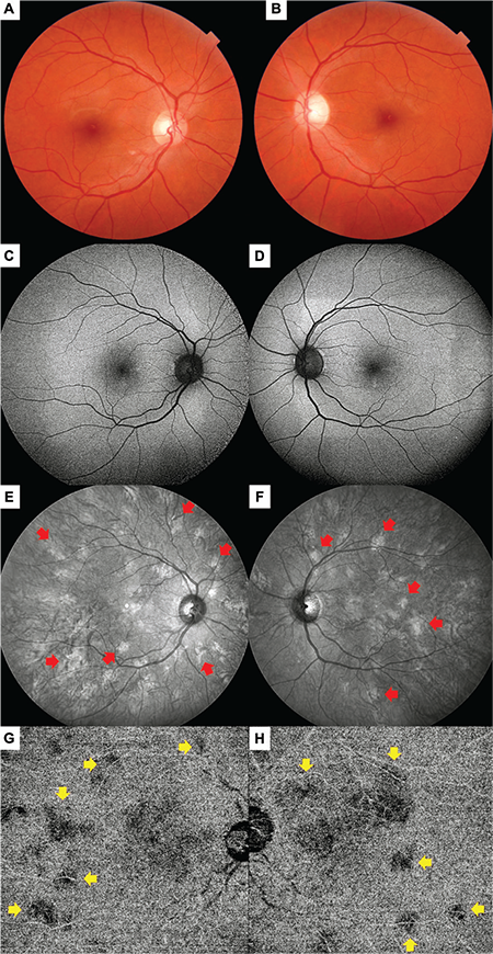
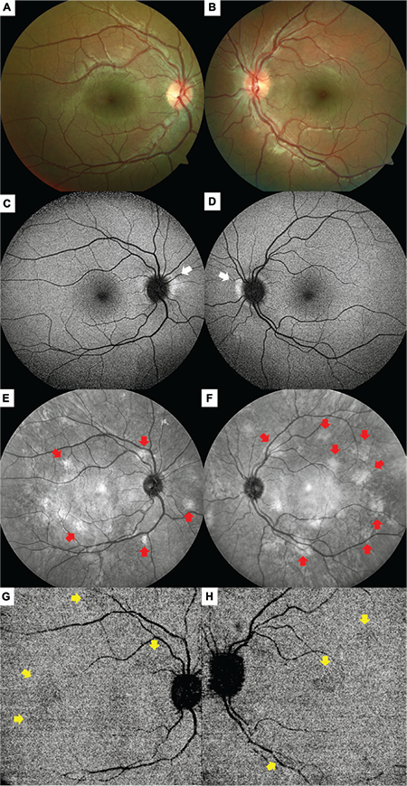
IR imaging revealed at least one choroidal nodule in 52 eyes (96.3%). One pediatric patient (3.7%) had bilateral Lisch nodules but no detectable choroidal nodule in either eye (patient 11, 11 years old). Adequate quality OCTA images were available for 43 eyes (79.6%). In 31 (72.1%) of these eyes, choroidal nodules were observed as areas of low reflectivity in the choriocapillaris layer.
Two (3.7%) of the 54 eyes included in the study had no choroidal nodules despite the presence of Lisch nodules. In contrast, 16 eyes (29.6%) had no Lisch nodules despite the presence of choroidal nodules. In the remaining 36 eyes (66.7%), both Lisch nodules and choroidal nodules were detected.
The multimodal imaging characteristics of patient 2 are shown in and , and those of patient 18 are presented in and as examples.
Discussion
Histopathological studies have revealed that the choroidal nodules observed in NF-1 are hamartomatous in character, consisting of ovoid bodies that contain Schwann cells which proliferate in an annular structure around axons, and may also include neural crest-derived melanocytes and ganglion cells., , These nodules are reported to impact choroidal blood flow, causing compression and thinning of the choriocapillaris and subsequently leading to choroidal and retinal thinning., These lesions also show structural similarities to the cutaneous neurofibromas and Lisch nodules observed in the iris.
Due to the high wavelength light used, IR imaging is a suitable modality for evaluating the ocular structures that lie beyond the RPE and is useful in detecting changes at the choroidal level. Although the choroidal nodules of NF-1 patients are not visible on fundoscopic examination, fundus autofluorescence imaging, or fluorescein angiography, they appear as bright, patchy areas in IR imaging and as hypocyanescent, patchy areas in the early phases of indocyanine green angiography., In addition, the bright, patchy areas observed on IR imaging were found to be areas of high flow in the deep choroidal segment of OCTA. In our study, choroidal nodules were observed in 31 (72.1%) of 43 eyes with adequate quality OCTA images, appearing as areas of flow deficit in the choriocapillaris layer. Although it is generally accepted that these nodules cannot be observed on fundoscopic examination, some authors have stated that the degree of pigmentation may vary and as a result, areas of hyperpigmentation in the fundus might be detectable in some cases.,
Studies in the literature that used IR imaging to examine choroidal nodules in NF-1 are summarized in ., , , , , , , , , , , , , , , , ,
Table 3. Studies evaluating the presence of choroidal nodules with infrared reflectance imaging in neurofibromatosis type 1
Authors (reference) | Number of patients/eyes, n | Age (years) mean ± SD | CN presence, n (%) | Remarks |
|---|---|---|---|---|
Yasunari et al.8 | 17 patients | 29.2±18.0 | 17 (100%) | - The bright patchy areas seen on infrared fundus examination and the hypofluorescent areas corresponding to these areas on indocyanine green angiography are likely of choroidal origin. - The high frequency of these abnormalities suggests that choroidal tissue is one of the most commonly affected structures in NF-1. |
Arigon et al.11 | 211 patients | 32.0±14.0 | 61 (28.9%) | - CN have a hamartomatous character and increase in incidence with age, similar to Lisch nodules. - CN are the most common ophthalmologic finding in NF-1 patients after Lisch nodules. - Red-free imaging is the most effective method for detecting CN. - CN may appear hypopigmented or hyperpigmented on ophthalmoscopic examination and hypofluorescent or hyperfluorescent on fluorescein angiography. |
Moramarco et al.13 | 249 patients | 33.0±17.1 | 238 (95.6%) | - Hyperpigmentation can be observed on fundoscopic examination in areas corresponding to the location of CN. - These areas tend to be located at the posterior pole because the area between the vascular arcades has thick choroidal tissue and is rich in melanocytes and neural cells. |
Abdolrahimzadeh et al.18 | 19 patients | 42.8±14.3 | 17 (89.4%) | - In NF-1 patients, CN may appear as a dome or placoid shape. - These lesions lead to changes in choroidal thickness and morphology. |
Vagge et al.19 | 78 patients | 8.1±3.5 | 54 (69.2%) | - CN are very common in children with NF-1. - CN can be visualized on IR imaging as bright patchy areas and are diagnostic. |
Cassiman et al.20 | 34 patients | 22.0 | 23 (68%) | - CN are easily detected with a confocal laser scanning ophthalmoscope. - CN may vary in prevalence and number according to different genotypic subtypes of NF-1 and aid in the differential diagnosis from Legius syndrome. - Detecting CN facilitates the diagnosis, especially in young patients without the fully developed clinical picture. |
Chilibeck et al.21 | 74 patients | Unspecified | 56 (75.7%)* | - CN detected by IR imaging are common in NF-1 and appear at an early age. - IR imaging can aid in the diagnosis of NF-1 in young children. |
Godinho et al.22 | 44 eyes | 16.4±7.3 | 28 (63.4%) | - The presence of CN correlates with the presence of central nervous system findings in NF-1 patients. |
de Rivas et al.23 | 30 patients | 57.0 | 25 (83.3%) | - CN, which can be detected on IR imaging, are common in NF-1 patients, similar to Lisch nodules, and are even more common than Lisch nodules when high myopic eyes are excluded. |
Goktas et al.24 | 19 patients | 8.6±3.2 | 15 (78.9) | - Choroidal abnormalities are common in NF-1 patients. - Choroidal abnormalities detectable by OCT and IR imaging can be used to diagnose NF-1. |
Viola et al.25 | 95 patients | 28.0±16.0 | 79 (82%) | - CN appear as bright, patchy areas on IR imaging and are common in NF-1. - IR imaging should be considered as a new diagnostic criterion in order to detect CN in NF-1 patients. |
Makino et al.26 | 10 patients | 20.6±13.2 | 9 (90%) | - Choroidal abnormalities in NF-1 patients increase with age and are correlated with the number of Lisch nodules. |
Vagge et al.27 | 31 patients | 22.7±13.7 | 24 (77.4%) | - Retinal thickness changes and CN are among the ocular symptoms described in NF-1 cases. - OCTA is an important technique in detecting the retinal and choroidal vascular flow changes that may occur early in NF-1. |
Moramarco et al.28 | 160 patients | 32.0±17.0 | 156 (97%) | - IR imaging is a non-invasive, sensitive, and reproducible in vivo imaging method that can be used to detect CN in NF-1 patients. - Choroidal changes are a diagnostic feature of NF-1. |
Estrela et al.29 | 41 patients | 11.8±3.3 | 39 (47.6%) | - Choroidal abnormalities are common in children with NF-1 and optic glioma but may not be detected in all cases. - Although choroidal abnormalities are a diagnostic criterion for NF-1, their presence and size have no impact on visual function. |
Touzé et al.30 | 141 patients | 8.6 | 97 (68.8%) | - With age, NF-1 patients show an increase in CN number and area on IR imaging. |
Flores Pimentel et al.31 | 94 patients | 10.3±4.2 | 60 (64%) | - In pediatric NF-1 patients, CN are more common than Lisch nodules, regardless of age and genetic confirmation. - Combining ophthalmological examination with IR imaging will be beneficial for early diagnosis in children. |
Parrozzani et al.32 | 119 patients | 8.3±4.5 | 72 (60.5%) | - Choroidal abnormalities can be regarded as a diagnostic criterion for the diagnosis of NF-1 in children. - The main advantage of this finding is that it aids early diagnosis, while the main drawback is that patient cooperation is required for its detection. |
Ahmadova et al. | 54 eyes | 22.3±14.6 | 52 (96.3%) | - CN are more common than Lisch nodules in NF-1 patients. - The earlier appearance of CN compared to Lisch nodules may facilitate early diagnosis in these patients. |
SD: Standard deviation, NF-1: Neurofibromatosis type 1, CN: Choroidal nodules, IR: Infrared reflectance
There are publications reporting that choroidal abnormalities, which can be detected up to 100% of NF-1 patients, are much more common than cafe au lait spots (98%) and Lisch nodules (41-68%) (classically considered the most common findings of the disease) and may actually be the most common sign of NF-1., Similar to cutaneous findings and Lisch nodules, choroidal findings are observed more frequently with older age, but they occur earlier than Lisch nodules. The prevalence of choroidal findings and Lisch nodules in pediatric cases has been reported to be 64-95% and 41-52%, respectively.,
Moreover, choroidal findings without Lisch nodules have been observed in 14-37% of NF-1 cases, while Lisch nodules without choroidal findings were observed in 2.5-16%., , In our series, choroidal nodules were observed in 29.6% of eyes without Lisch nodules, whereas 3.7% of the eyes had only Lisch nodules.
Study Limitations
The main limitations of this study are that it was retrospective and single-centered with a relatively small number of patients. In addition, assessments that can indicate functional status, such as visual acuity and visual field, were not included in our study. However, the reason for this is that most previous publications examining choroidal nodules in NF cases have stated that these findings are asymptomatic and do not cause functional problems. Furthermore, as the OCTA images in our study were recorded for the purpose of evaluating for choroidal nodules, 12x12-mm images were obtained in order to evaluate the largest possible area. However, due to the features of the OCTA device we used, we could not obtain data on quantitative parameters such as vascular density and the foveal avascular zone with 12x12-mm images. Considering that choroidal nodules can lead to vascular alterations, especially at the choriocapillaris level, this can also be considered a limitation.
Conclusion
As NF-1 can cause ophthalmological findings, patients often present or are referred to ophthalmology clinics, both through outpatient clinic admissions and consultations by other medical disciplines. Therefore, it is important for ophthalmologists to recognize the ocular findings of NF-1. Although the presence of Lisch nodules can be evaluated by biomicroscopic examination, choroidal nodules can be observed much more frequently in these patients, as in our series, and can be easily detected using IR imaging, which is a rapid and non-invasive method. The presence of choroidal nodules before the appearance of Lisch nodules may facilitate early diagnosis in these patients.
Footnotes
Ethics
Ethics Committee Approval: Dokuz Eylül University Local Ethics Committee (approval number: 2023/10-01, date: 29.03.2023).
Informed Consent: Retrospective study.
Authorship Contributions
Concept: M.K., S.K., A.O.S., Design: M.K., E.H., Data Collection or Processing: N.A., M.K., E.H., Analysis or Interpretation: M.K., S.K., A.O.S., Literature Search: N.A., M.K., S.K., Writing: N.A., M.K., A.O.S.
Conflict of Interest
Conflict of Interest: No conflict of interest was declared by the authors.
Financial Disclosure
Financial Disclosure: The authors declared that this study received no financial support.
References
Abdolrahimzadeh S, Plateroti AM, Recupero SM, Lambiase A. An Update on the Ophthalmologic Features in the Phakomatoses. J Ophthalmol. 2016;2016:3043026.
Ratner N, Miller SJ. A RASopathy gene commonly mutated in cancer: the neurofibromatosis type 1 tumour suppressor. Nat Rev Cancer. 2015;15:290-301.
Bajaj A, Li QF, Zheng Q, Pumiglia K. Loss of NF1 expression in human endothelial cells promotes autonomous proliferation and altered vascular morphogenesis. PLoS One. 2012;7:e49222.
Saatci AO, Saylam GS, Yasti ZO, Söylev M, Saatci I, Kavukçu S, Memişoğlu B. Neurofibromatosis type I and unilateral ophthalmic artery occlusion. Ophthalmic Genet. 1998;19:87-91.
Özdemir Zeydanlı E, Özdek Ş. Neurofibromatosis Type 1 Vasculopathy Presenting as Branch Retinal Vein Occlusion: Case Report and Review of the Literature. Turk J Ophthalmol. 2023;53:390-394.
No authors listed. Neurofibromatosis. Conference statement. National Institutes of Health Consensus Development Conference. Arch Neurol. 1988;45:575-578.
Legius E, Messiaen L, Wolkenstein P, Pancza P, Avery RA, Berman Y, Blakeley J, Babovic-Vuksanovic D, Cunha KS, Ferner R, Fisher MJ, Friedman JM, Gutmann DH, Kehrer-Sawatzki H, Korf BR, Mautner VF, Peltonen S, Rauen KA, Riccardi V, Schorry E, Stemmer-Rachamimov A, Stevenson DA, Tadini G, Ullrich NJ, Viskochil D, Wimmer K, Yohay K; International Consensus Group on Neurofibromatosis Diagnostic Criteria (I-NF-DC); Huson SM, Evans DG, Plotkin SR. Revised diagnostic criteria for neurofibromatosis type 1 and Legius syndrome: an international consensus recommendation. Genet Med. 2021;23:1506-1513.
Yasunari T, Shiraki K, Hattori H, Miki T. Frequency of choroidal abnormalities in neurofibromatosis type 1. Lancet. 2000;356:988-992.
Safi A, Borruat FX. Yasunari Nodules: A New Sensitive and Specific Marker of Neurofibromatosis Type 1, Readily Detectable by Ophthalmologists. Klin Monbl Augenheilkd. 2019;236:480-482.
Lubs ML, Bauer MS, Formas ME, Djokic B. Lisch nodules in neurofibromatosis type 1. N Engl J Med. 1991;324:1264-1266.
Arigon V, Binaghi M, Sabouret C, Zeller J, Revuz J, Soubrane G, Wolkenstein P. Usefulness of systematic ophthalmologic investigations in neurofibromatosis 1: a cross-sectional study of 211 patients. Eur J Ophthalmol. 2002;12:413-418.
Kurosawa A, Kurosawa H. Ovoid bodies in choroidal neurofibromatosis. Arch Ophthalmol. 1982;100:1939-1941.
Moramarco A, Mallone F, Sacchetti M, Lucchino L, Miraglia E, Roberti V, Lambiase A, Giustini S. Hyperpigmented spots at fundus examination: a new ocular sign in Neurofibromatosis Type I. Orphanet J Rare Dis. 2021;16:147.
Cassiman C, Casteels I, Stalmans P, Legius E, Jacob J. Optical Coherence Tomography Angiography of Retinal Microvascular Changes Overlying Choroidal Nodules in Neurofibromatosis Type 1. Case Rep Ophthalmol. 2017;8:214-220.
Formisano M, Lodesani M, Rullo D, Fabrizio L, Guglielmelli F, Scuderi G. Unusual case of enlarged choroidal vessels in neurofibromatosis type 1 observed with near-infrared reflectance and spectral domain optical coherence tomography. Clin Ter. 2021;172:507-510.
Richetta A, Giustini S, Recupero SM, Pezza M, Carlomagno V, Amoruso G, Calvieri S. Lisch nodules of the iris in neurofibromatosis type 1. J Eur Acad Dermatol Venereol. 2004;18:342-344.
Kumar V, Singh S. Multimodal imaging of choroidal nodules in neurofibromatosis type-1. Indian J Ophthalmol. 2018;66:586-588.
Abdolrahimzadeh S, Felli L, Plateroti R, Plateroti AM, Giustini S, Calvieri S, Recupero SM. Morphologic and vasculature features of the choroid and associated choroid-retinal thickness alterations in neurofibromatosis type 1. Br J Ophthalmol. 2015;99:789-793.
Vagge A, Camicione P, Capris C, Sburlati C, Panarello S, Calevo MG, Traverso CE, Capris P. Choroidal abnormalities in neurofibromatosis type 1 detected by near-infrared reflectance imaging in paediatric population. Acta Ophthalmol. 2015;93:667-671.
Cassiman C, Casteels I, Jacob J, Plasschaert E, Brems H, Dubron K, Keer KV, Legius E. Choroidal abnormalities in café-au-lait syndromes: a new differential diagnostic tool? Clin Genet. 2017;91:529-535.
Chilibeck CM, Shah S, Russell HC, Vincent AL. The presence and progression of choroidal neurofibromas in a predominantly pediatric population with neurofibromatosis type-1. Ophthalmic Genet. 2021;42:223-239.
Godinho G, Esteves-Leandro J, Alves G, Madeira C, Faria O, Brandão E, Magalhães A, Falcão-Reis F, Penas S. Correlation Between Ophthalmologic and Neuroradiologic Findings in Type 1 Neurofibromatosis. J Neuroophthalmol. 2022;42:101-107.
de Rivas MO, Gabás JM, Cabeza MÁT, Floría OE, Latorre RH, Moscarda EN, Clavería JA, Rivasés GP, Puyuelo JA. Choroidal Hyperreflective Nodules Detected by Infrared Reflectance Images Are a Diagnostic Criterion for Neurofibromatosis Type 1 Patients Excluding Those with High Myopia. Diagnostics (Basel). 2023;13:1348.
Goktas S, Sakarya Y, Ozcimen M, Alpfidan I, Uzun M, Sakarya R, Yarbag A. Frequency of choroidal abnormalities in pediatric patients with neurofibromatosis type 1. J Pediatr Ophthalmol Strabismus. 2014;51:204-208.
Viola F, Villani E, Natacci F, Selicorni A, Melloni G, Vezzola D, Barteselli G, Mapelli C, Pirondini C, Ratiglia R. Choroidal abnormalities detected by near-infrared reflectance imaging as a new diagnostic criterion for neurofibromatosis 1. Ophthalmology. 2012;119:369-375.
Makino S, Tampo H, Arai Y, Obata H. Correlations between choroidal abnormalities, Lisch nodules, and age in patients with neurofibromatosis type 1. Clin Ophthalmol. 2014;8:165-168.
Vagge A, Corazza P, Ferro Desideri L, Camicione P, Agosto G, Vagge R, Maria Grazia C, Carnevali A, Giannaccare G, Nicolò M, Traverso CE. Ocular biometric parameters changes and choroidal vascular abnormalities in patients with neurofibromatosis type 1 evaluated by OCT-A. PLoS One. 2021;16:e0251098.
Moramarco A, Giustini S, Nofroni I, Mallone F, Miraglia E, Iacovino C, Calvieri S, Lambiase A. Near-infrared imaging: an in vivo, non-invasive diagnostic tool in neurofibromatosis type 1. Graefes Arch Clin Exp Ophthalmol. 2018;256:307-311.
Estrela T, Truong S, Garcia A, He J, Ying GS, Devakandan K, Reginald YA, Fisher MJ, Liu GT, Ullrich NJ, Avery RA, Heidary G. The Relationship Between Choroidal Abnormalities and Visual Outcomes in Pediatric Patients With NF1-Associated Optic Pathway Gliomas. J Neuroophthalmol. 2024;44:5-9.
Touzé R, Manassero A, Bremond-Gignac D, Robert MP. Long-term follow-up of choroidal abnormalities in children with neurofibromatosis type 1. Clin Exp Ophthalmol. 2021;49:516-519.
Flores Pimentel M, Heath A, Wan MJ, Hussein R, Leahy KE, MacDonald H, Tavares E, VandenHoven C, MacNeill K, Kannu P, Parkin PC, Heon E, Reginald A, Vincent A. Prevalence of Choroidal Abnormalities and Lisch Nodules in Children Meeting Clinical and Molecular Diagnosis of Neurofibromatosis Type 1. Transl Vis Sci Technol. 2022;11:10.
Parrozzani R, Clementi M, Frizziero L, Miglionico G, Perrini P, Cavarzeran F, Kotsafti O, Comacchio F, Trevisson E, Convento E, Fusetti S, Midena E. In Vivo Detection of Choroidal Abnormalities Related to NF1: Feasibility and Comparison With Standard NIH Diagnostic Criteria in Pediatric Patients. Invest Ophthalmol Vis Sci. 2015;56:6036-6042.
Mallone F, Lucchino L, Giustini S, Lambiase A, Moramarco A. An update on choroidal abnormalities and retinal microvascular changes in neurofibromatosis type 1. Orphanet J Rare Dis. 2022;17:223.

