

Dear Editor,
Ocular blunt trauma can lead to cyclodialysis, which may resolve spontaneously or respond to conservative management. However, interventional approaches may be required when the cleft is refractory to medical therapy., Surgical closure of the cleft is generally effective at restoring intraocular pressure (IOP), but the occurrence of postoperative complications (such as severe IOP spikes) sometimes necessitates additional glaucoma surgery.,
This letter reports an unusual case of long-standing ocular hypotony due to a chronic cyclodialysis cleft. The case was further complicated by the development of severe, medically uncontrolled ocular hypertension following surgical cleft closure, which was managed by suture revision.
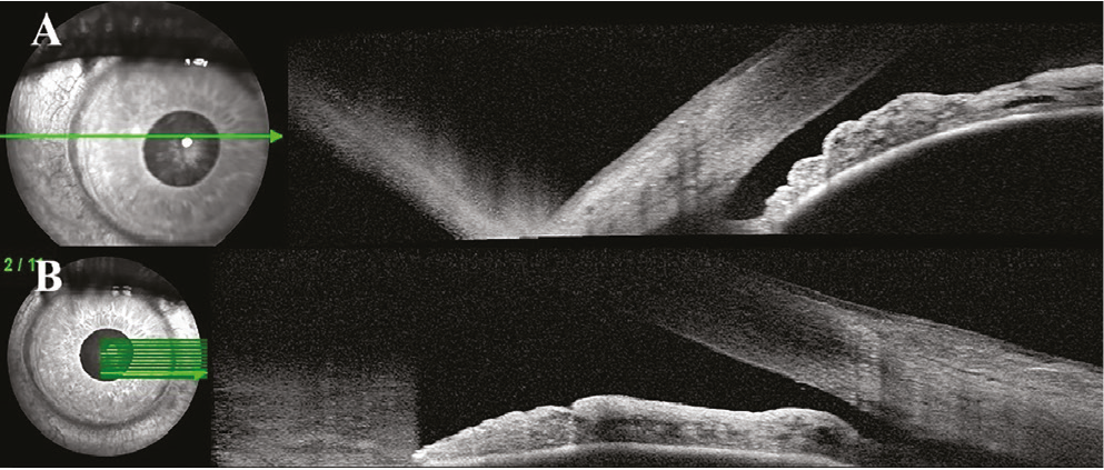
A 52-year-old man presented with decreased vision in his right eye and history of a severe motor vehicle accident 5 years earlier. On examination, best corrected visual acuity (BCVA) was hand motion in the right eye. IOP was 5 mmHg in the right eye and 15 mmHg in the left eye. Anterior segment examination of the right eye revealed an intumescent cataract and a markedly shallow anterior chamber. Fundus examination could not be performed due to media opacity. B-scan ultrasonography revealed no retinal or choroidal detachment. However, the axial length (AL) of the right eye was approximately 2 mm shorter than that of the left (22.15 mm vs. 23.99 mm). Gonioscopic evaluation was limited due to hypotony; the angle structures appeared indistinct and irregular secondary to trauma. Ultrasound biomicroscopy was recommended but declined by the patient. Anterior segment optical coherence tomography was subsequently performed; 360-degree scanning revealed a cyclodialysis cleft at the nasal angle and a blunt angle configuration in the inferior and temporal quadrants ().
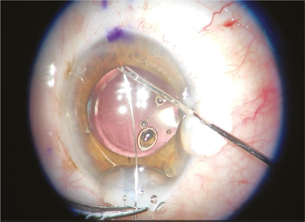
Phacoemulsification, cleft closure using the cross-chamber cyclopexy technique, and capsular tension ring (CTR) implantation in the sulcus were performed. Following uneventful cataract removal, an approximately 1-clock-hour cyclodialysis cleft (from 3 to 4 o’clock) was identified at the nasal angle using a Swan-Jacob gonioprism lens. The cleft area was identified and marked on the sclera. A double-armed 10-0 polypropylene suture was passed into the anterior chamber, exiting 1.5 mm posterior to the limbus at both outer edges of the cleft (). The conjunctiva was closed after the suture was tightened and tied over the sclera. Prior to viscoelastic removal, a Morcher type 14A CTR was placed in the ciliary sulcus to ensure that no additional cyclodialysis areas were missed.
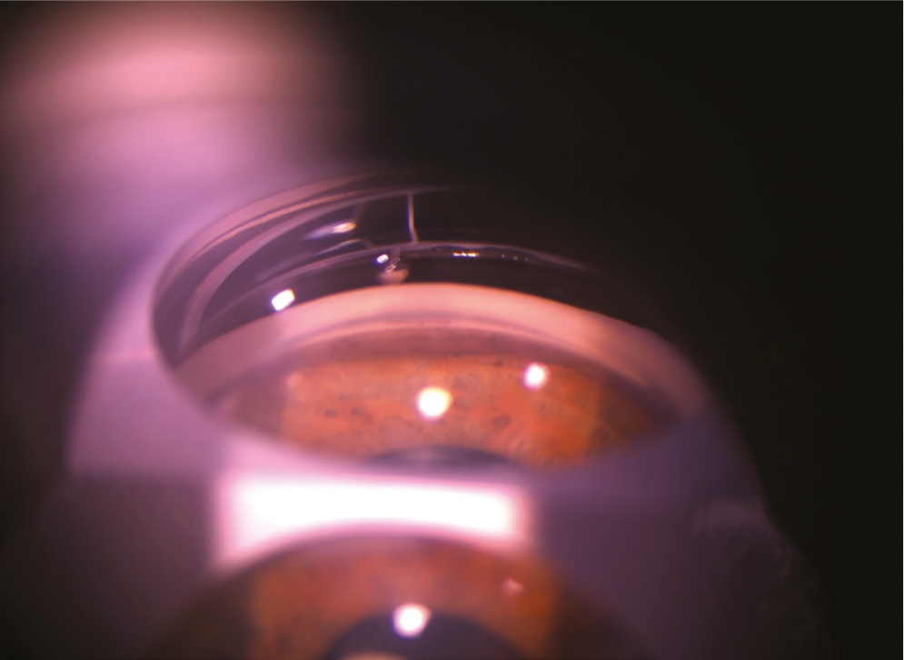
IOP was 55 mmHg on postoperative day 1. The patient was managed with maximum topical antiglaucomatous therapy. Systemic treatment included oral acetazolamide (Diazomid®, Sanofi Health Products, İstanbul, Türkiye) thrice daily and intravenous mannitol (5 mL/kg, Polifarma Pharmaceuticals, İstanbul, Türkiye) twice daily. No improvement in IOP was observed until the end of postoperative day 5, with a lowest recorded value of 45 mmHg. Gonioscopy revealed a closed cleft with a broadly and irregularly widened ciliary body band and indistinct angle structures, consistent with angle recession and trabecular damage (). Due to severe, medically uncontrolled ocular pain and nausea associated with persistently elevated IOP, the 10-0 polypropylene suture used for cyclodialysis cleft closure was removed, given that a CTR had also been implanted in the sulcus during the initial surgery.
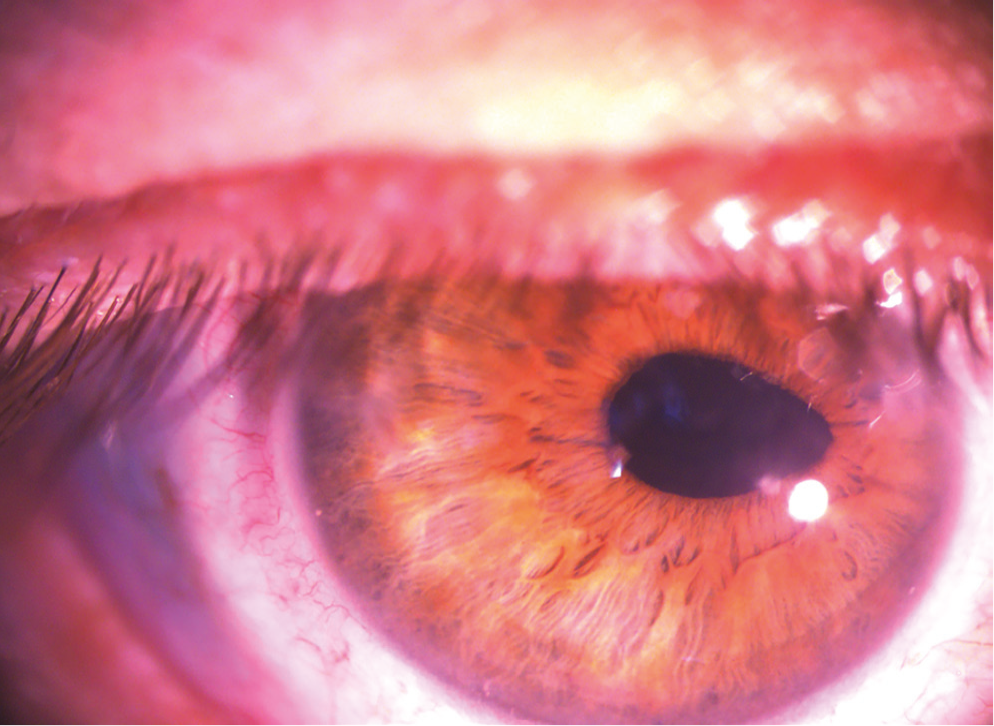
Following suture removal, the IOP rapidly decreased to 6 mmHg. At 12 weeks, BCVA improved to 0.6 (Snellen decimal), with an IOP of 14 mmHg in the right eye. However, a localized pupillary retraction was evident on the nasal side (). Optical coherence tomography showed resolution of hypotony maculopathy (). However, the AL was similar to the preoperative measurement (22.22 mm) and gonioscopy revealed a partially open cyclodialysis cleft smaller than the preoperative cleft area ().
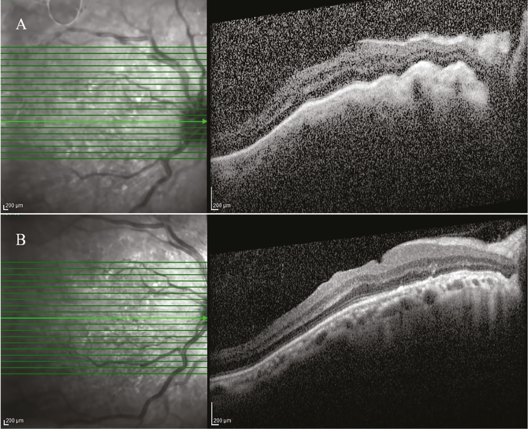
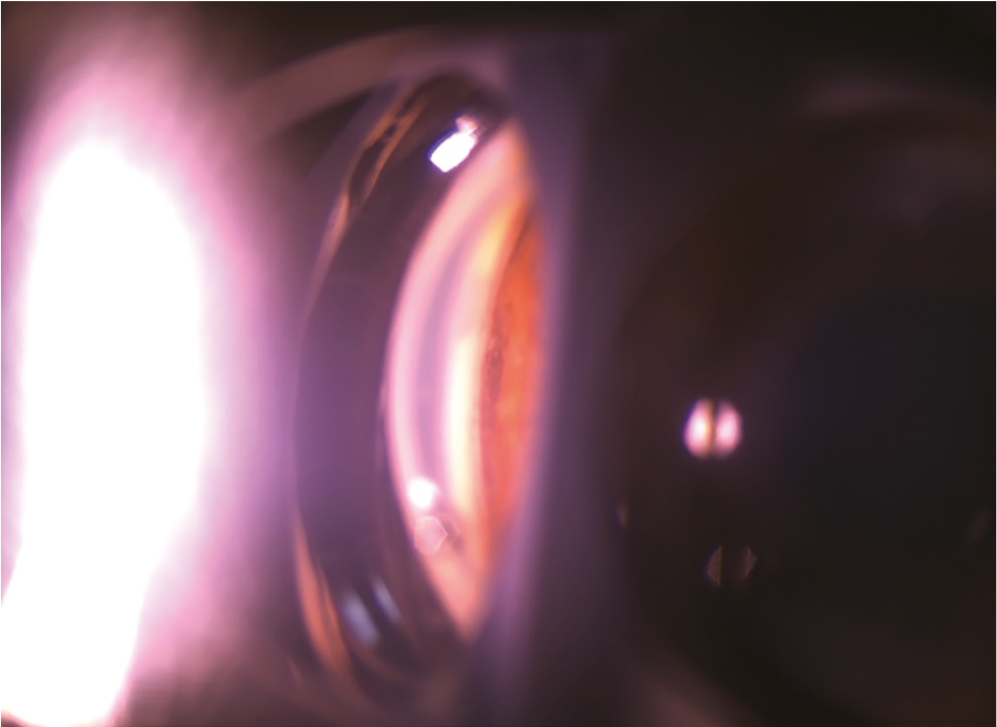
A notable postoperative event in this case was the rapid and severe IOP elevation to 55 mmHg, which was unresponsive to intensive medical therapy. Hypertensive episodes following cyclodialysis cleft closures are well documented, with reported incidence ranging from 48% to 84%., This phenomenon is attributable to the abrupt cessation of abnormal aqueous outflow through the cleft and the subsequent reactivation of a previously compromised trabecular meshwork which may have become functionally atrophic during prolonged hypotony.
Post-cleft closure IOP spikes may be refractory to medical therapy and necessitate additional glaucoma surgery., Ohtani et al. reported a case in which Ahmed glaucoma valve implantation was performed 14 days after direct internal cyclopexy due to persistent postoperative IOP elevation. In another study, both eyes required additional surgery following surgical cleft closure. Similarly, this patient experienced medically uncontrolled elevated IOP with severe pain and nausea, necessitating further intervention. Additionally, the patient’s ocular features (including the angle appearance consistent with recession and trabecular damage) and the prolonged hypotony, which can lead to trabecular dysfunction, warranted surgical revision instead of continued follow-up with antiglaucomatous medications. Suture removal (rather than a second IOP-lowering procedure) was performed as the sulcus CTR could facilitate cleft closure over time, while suture removal alone could effectively reduce IOP.
Trabecular dysfunction sustained by ocular hypotony is suggested to reverse following cleft closure, potentially contributing to gradual normalization of the IOP. However, we consider this unlikely in patients with chronic hypotony lasting several years. In line with this view, Agrawal and Shah hypothesized that microscopic aqueous drainage into the suprachoroidal space may persist despite clinically evident closure of the cyclodialysis cleft. In this case, final gonioscopy revealed a partially open cyclodialysis cleft (<0.5 clock hours) smaller than the preoperative cleft size. Therefore, we believe that the equilibrium between aqueous production and outflow is maintained by the residual cleft area rather than trabecular function recovery.
Based on this patient’s experience, we recommend the use of adjustable sutures in incisional cyclodialysis closure surgeries. This technique, which permits aqueous drainage after loosening, may help reduce postoperative uncontrolled IOP spikes and the need for secondary glaucoma procedures.
Footnotes
Ethics
Informed Consent: Written informed consent was obtained from the patient.
Authorship Contributions
Surgical and Medical Practices: A.M.K., Concept: A.M.K., Design: A.M.K., Data Collection or Processing: A.M.K., B.E.A., Analysis or Interpretation: A.M.K., Literature Search: A.M.K., Writing: A.M.K.
Conflict of Interest
Conflict of Interest: No conflict of interest was declared by the authors.
Financial Disclosure
Financial Disclosure: The authors declared that this study received no financial support.
References
González-Martín-Moro J, Contreras-Martín I, Muñoz-Negrete FJ, Gómez-Sanz F, Zarallo-Gallardo J. Cyclodialysis: an update. Int Ophthalmol. 2017;37:441-457.
Leuzinger-Dias M, Lima-Fontes M, Oliveira-Ferreira C, Macedo JP, Falcão-Reis F, Freitas-da-Costa P, Benevides-Melo A. Traumatic cyclodialysis cleft treatment combined with cataract surgery: an original triple procedure. Ophthalmol Ther. 2021;10:1171-1179.
Delgado MF, Daniels S, Pascal S, Dickens CJ. Hypotony maculopathy: improvement of visual acuity after 7 years. Am J Ophthalmol. 2001;132:931-933.
Ohtani H, Harano A, Ichioka S, Shimada A, Iida M, Murakami K, Ida C, Tanito M. A case of long-term undiagnosed cyclodialysis cleft following tanito microhook trabeculotomy. Cureus. 2025;17:e77516.
Ioannidis AS, Bunce C, Barton K. The evaluation and surgical management of cyclodialysis clefts that have failed to respond to conservative management. Br J Ophthalmol. 2014;98:544-549.
Leen MM, Mills RP. Low postoperative intraocular pressure. In: Spaeth G, ed. Ophthalmic Surgery: Principles and Practice. Philadelphia, PA: Saunders; 2003:380-387.
Agrawal P, Shah P. Long-term outcomes following the surgical repair of traumatic cyclodialysis clefts. Eye (Lond). 2013;27:1347-1352.
Küchle M, Naumann GO. Direct cyclopexy for traumatic cyclodialysis with persisting hypotony. Report in 29 consecutive patients. Ophthalmology. 1995;102:322-333.
Hwang JM, Ahn K, Kim C, Park KA, Kee C. Ultrasonic biomicroscopic evaluation of cyclodialysis before and after direct cyclopexy. Arch Ophthalmol. 2008;126:1222-1225.

