

Dear Editor,
Corneal ulcers are vision-threatening emergencies. Bacterial pathogens such as Staphylococcus, Streptococcus, and Pseudomonas are most frequently implicated in microbial keratitis. The integrity of the corneal epithelium is critical in preventing infections, as it acts as a physical and immunological barrier.
Enterobacter cloacae is a Gram-negative rod in the Enterobacteriaceae family, commonly present in the environment and human gut microbiota. Although increasingly linked to nosocomial infections, ocular involvement remains rare. The prevalence of E. cloacae keratitis ranges from 0.13% to 15.3%., Most documented cases involve penetrating keratoplasty (PK), graft failure, chronic corneal edema, or topical corticosteroid use., , ,
Dry eye disease (DED) is a multifactorial disorder characterized by tear film instability, ocular surface inflammation, and neurosensory abnormalities. Severe DED leads to disruption of the corneal epithelial barrier, impaired wound healing, and reduced resistance to pathogens. Topical corticosteroids or calcineurin inhibitors may exacerbate these effects.
We present a rare case of E. cloacae keratitis that highlights the predisposing role of severe dry eye and topical corticosteroids and briefly review previously reported cases to contextualize the risk factors associated with this opportunistic infection.
A 77-year-old woman who did not use contact lenses presented with a 1-week history of pain, redness, and decreased vision in the right eye. Her best corrected visual acuity (BCVA) had declined from 20/25 to 20/32. She had a 3-year history of severe DED, with Schirmer test values of 4 and 3 mm, tear break-up time of 3 seconds, and grade II punctate keratitis in both eyes, together with meibomian gland dysfunction. No systemic predisposing conditions or previous ocular surgery were reported. The patient maintained adequate hygiene.
Treatment included warm compresses, lid hygiene, artificial tears (Thealoz Duo®; Laboratoires Théa, Clermont-Ferrand, France), and tear ointment (VitA-POS®; URSAPHARM Arzneimittel GmbH, Saarbrücken, Germany) at bedtime. Topical corticosteroid (dexamethasone 1 mg/mL, preservative-free; Dexafree®, Laboratoires Théa, Clermont-Ferrand, France) had been started 6 weeks earlier and was being tapered with a dose reduction every 10 days. At presentation, she was using fluorometholone 1 mg/mL (FML®, Allergan Pharmaceuticals, Westport, Ireland) once daily and cyclosporine A 0.05% eye drops (hospital-compounded formulation, Barraquer Ophthalmology Center Pharmacy, Barcelona, Spain) twice daily.
Slit-lamp examination showed mucous discharge, conjunctival hyperemia, and a 2-mm corneal ulcer with dense stromal infiltrate and surrounding calcium deposits in the inferior midperiphery (5 o’clock). No keratic precipitates were observed ().
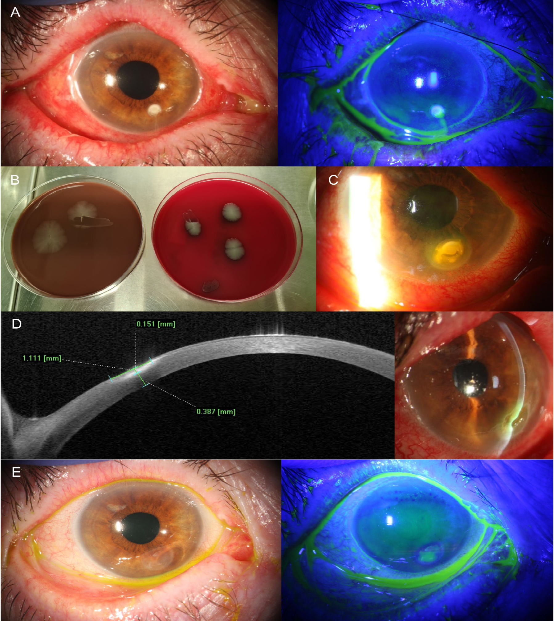
Suspecting microbial keratitis, corneal scrapings were obtained for culture. Empirical treatment was begun with fortified cefazolin 50 mg/mL and amikacin 20 mg/mL eye drops (hospital-compounded formulation, Barraquer Ophthalmology Center Pharmacy, Barcelona, Spain) applied hourly while awake and ciprofloxacin 3 mg/g ointment (Oftacilox®, NTC Srl, Milan, Italy) at bedtime. Fluorometholone and cyclosporine A were discontinued.
Corneal cultures grew E. cloacae () sensitive to amikacin, ciprofloxacin, and ceftazidime. After 1 week of treatment, the ulcer was controlled and the epithelial defect was closed. Thus, fortified cefazolin and amikacin were discontinued, and treatment was switched to ceftazidime 50 mg/mL (hospital-compounded formulation, Barraquer Ophthalmology Center Pharmacy, Barcelona, Spain) 5 times daily to reduce toxicity to the ocular surface, maintaining ciprofloxacin ointment every 12 hours. Medroxyprogesterone 20 mg/mL eye drops (hospital-compounded formulation, Barraquer Ophthalmology Center Pharmacy, Barcelona, Spain) were added to control inflammation.
After 1 month, infection was controlled but the calcium deposit enlarged (), causing a new epithelial defect. Conservative management with daily fluorometholone, tobramycin 3 mg/g ointment (Tobrex®, Novartis Pharma, Barcelona, Spain), and an eye patch was started. As no improvement occurred after 3 days, corneal curettage was performed, leaving 387 µm of residual stroma on anterior segment tomography (). The same regimen achieved complete epithelial closure within 1 week.
Maintenance treatment for severe DED was then resumed, including 50% autologous serum drops hourly, fluorometholone 1 mg/mL once daily, and cyclosporine A 0.05% twice daily to control chronic ocular surface inflammation.
At 7-month follow-up, stromal thinning and corneal haze persisted (), but BCVA improved to 20/25. Maintenance therapy continued with 6-month follow-up.
E. cloacae keratitis is rare; only 13 cases have been reported (). Most occurred in patients with a history of PK, graft failure, or prolonged topical corticosteroid use, often associated with corneal bullae, irregular ocular surface, or neurotrophic keratopathy. Gross and Meyer noted reduced tear production as a possible factor. Rajarajan et al. reviewed 7,787 infectious keratitis cases and identified E. cloacae in only 10 (0.13%): 9 following PK and 1 associated with bullous keratopathy. They suggested that epithelial bullae and an irregular corneal surface from graft failure may compromise the epithelial barrier, particularly when combined with long-term corticosteroid use. The most recent case, reported by Al Rasheed et al., occurred after corneal cross-linking with epithelial debridement, further emphasizing epithelial disruption as a major predisposing factor. Additionally, Feizi et al. reported E. cloacae contamination in 7.02% of therapeutic bandage contact lenses, suggesting possible environmental exposure.
Table 1. Reported cases of Enterobacter cloacae keratitis in the literature and proposed risk factors
Cases | Gender/age | Previous surgeries | Risk factors | Presenting VA | Ulcer size and morphology | Sensitive antibiotic | Final VA | Outcome/duration (d) |
|---|---|---|---|---|---|---|---|---|
Gross and Meyer (1985) | F/70 | -Extracapsular cataract extraction -PK for pseudophakic bullous keratopathy | -Chronic edema after graft rejection -Topical corticosteroid therapy -Tear insufficiency -Poor toilet hygiene and inadequate hand washing | HM at 15 cm | 3.0x1.2 mm Dense, white stromal infiltrate | Gentamicin and sulfacetamide | NM | Residual edema secondary to previous graft rejection/7 |
Sharma et al. (2020) | F/50 | PK | NM except for surgery | HM close to face | Multifocal infiltrates of varying size | Ceftazidime | NM | Regraft/NM |
Rajarajan et al. (2021) (Case 1) | F/62 | PK | -Failed PK -Corneal bullae and irregular ocular surface -Topical corticosteroid therapy | CF at 10 cm | 6 mm2 Patchy yellow | Ciprofloxacin | CF at 10 cm | Resolved/45 |
Rajarajan et al. (Case 2) | M/45 | PK | -PK -Corneal bullae and irregular ocular surface -Topical corticosteroid therapy -Neurotrophic keratopathy | CF at 10 cm | 2.7 mm2 Dense white | Ofloxacin | CF at 10 cm | Resolved/30 |
Rajarajan et al. (Case 3) | M/41 | PK | -Failed PK -Loose sutures -Topical corticosteroid therapy -Neurotrophic keratopathy | 20/50 | 2.7 mm2 Patchy white | Gentamicin, imipenem and chloramphenicol | 20/100 | Resolved/40 |
Rajarajan et al. (Case 4) | M/58 | PK | -Failed PK -Corneal bullae and irregular ocular surface -Topical corticosteroid therapy -Neurotrophic keratopathy | CF at 10 cm | 1 mm2 Patchy white | Ofloxacin | CF at 10 cm | Resolved/9 |
Rajarajan et al. (Case 5) | M/55 | PK | -Failed PK -Corneal bullae and irregular ocular surface -Topical corticosteroid therapy | HM | 64 mm2 Dense yellow | Amikacin | HM | Graft edema/NM |
Rajarajan et al. (Case 6) | F/70 | PK | CF at 10 cm | 20.8 mm2 Dense yellow | Chloramphenicol | NLP | Phthisis/120 | |
Rajarajan et al. (Case 7) | M/50 | PK | -Failed PK -Topical corticosteroid therapy | Light perception | 27.9 mm2 Dense yellow | Gentamicin | HM | Scar/110 |
Rajarajan et al. (Case 8a) | M/50 | PK | -Failed PK -Corneal bullae and irregular ocular surface -Topical corticosteroid therapy | 20/400 | 27.44 mm2 Dense yellow | Azithromycin | CF at 10 cm | Regraft/60 |
Rajarajan et al. (Case 8b) | M/50 | PK | CF at 10 cm | 9.5 mm2 Dense yellow | Azithromycin | CF at 10 cm | Failed PK/45 | |
Rajarajan et al. (Case 9) | M/61 | NM | -Corneal bullae and irregular ocular surface -Topical corticosteroid therapy | 20/600 | 5.25 mm2 Patchy white | Gatifloxacin | 20/60 | Scar/45 |
Al Rasheed et al. (2023) | F/19 | Corneal cross-linking for keratoconus | -Non-adherence to post-procedure medications -Corneal epithelium debridement | 20/200 | 7.8 mm2 Ring-shaped | Amikacin and moxifloxacin | 20/40 | Stromal haze and subepithelial scar/30 |
Cintas et al. (present case) | F/77 | None | -Severe DED -Topical corticosteroid therapy | 20/32 | 2 mm2 Dense white with calcium deposits | Amikacin, ciprofloxacin and ceftazidime | 20/25 | Stromal thinning and corneal haze/70 |
CF: Counting fingers, d: Days, DED: Dry eye disease, F: Female, HM: Hand motion, M: Male, NLP: No light perception, NM: Not mentioned, PK: Penetrating keratoplasty, VA: Visual acuity
In our case, the absence of prior surgery, corneal decompensation, systemic disease, or contact lens use suggests that alternative risk factors must be considered. Severe DED is widely recognized to compromise epithelial integrity, impair tear film antimicrobial defense, and reduce corneal sensitivity, factors that collectively reduce ocular immunity.
Interestingly, as shown in , no previously reported case of E. cloacae keratitis identified corticosteroid use as the sole risk factor. In all cases, topical corticosteroids were associated with epithelial barrier disruption such as post-surgical status, corneal bullae, graft failure, or chronic surface disease. This reinforces the idea that both epithelial compromise and local immunosuppression are required to trigger opportunistic keratitis.
Corticosteroids, while reducing ocular surface inflammation, further impair local defense mechanisms and increase susceptibility to opportunistic infections in eyes with already compromised epithelial integrity. Our patient was using fluorometholone at presentation. Following TFOS DEWS III, medroxyprogesterone and autologous serum were used as corticosteroid-sparing therapy. Both provide regenerative and anti-inflammatory effects without relevant immunosuppression, making them appropriate for high-risk corneas.
An additional complication was the formation of a calcium deposit at the ulcer site, delaying epithelial healing and requiring curettage. This was likely related to phosphate-containing fluorometholone (FML, Allergan). In compromised ocular surfaces, phosphate buffers may precipitate with calcium in the tear film, forming hydroxyapatite crystals. Popiela and Hawksworth noted this calcification can occur regardless of dosage or duration and advised avoiding phosphate-based drops in chronic ocular surface disease. This case highlights the need to critically evaluate the use of corticosteroids in DED.
In conclusion, this case suggests that severe DED and topical immunosuppression may suffice to trigger infection by an opportunistic pathogen such as E. cloacae, even without classical risk factors like PK or graft failure. Evidence indicates that both epithelial disruption and corticosteroid use are usually required for the development of E. cloacae keratitis. Close monitoring of severe DED patients may prevent such infections.
Footnotes
Ethics
Informed Consent: Written informed consent was obtained from the patient for publication of the case and accompanying images.
Authorship Contributions
Surgical and Medical Practices: V.C., Concept: N.C., E.R.P., G.J., V.C., Design: N.C., E.R.P., G.J., V.C., Data Collection or Processing: V.C., Analysis or Interpretation: N.C., E.R.P., G.J., V.C., Literature Search: N.C., E.R.P., G.J., V.C., Writing: N.C., E.R.P.
Conflict of Interest
Conflict of Interest: No conflict of interest was declared by the authors.
Financial Disclosure
Financial Disclosure: The authors declared that this study received no financial support.
References
Ting DSJ, Ho CS, Deshmukh R, Said DG, Dua HS. Infectious keratitis: an update on epidemiology, causative microorganisms, risk factors, and antimicrobial resistance. Eye (Lond). 2021;35:1084-1101. Erratum in: Eye (Lond). 2021;35:2908.
Davin-Regli A, Lavigne JP, Pagès JM. Enterobacter spp.: update on taxonomy, clinical aspects, and emerging antimicrobial resistance. Clin Microbiol Rev. 2019;32:e00002-e00019.
Rajarajan M, Madduri B, Mohan N, Fernandes M. Enterobacter cloacae keratitis: clinicomicrobiological profiles, risk factors, and outcomes. Cornea. 2022;41:1110-1115.
Gross ND, Meyer RF. Enterobacter cloacae ulceration in a failed corneal graft: a case report. Br J Ophthalmol. 1985;69:542-544.
Sharma DR. Corneal infection by Enterobacter cloacae post penetrating keratoplasty. JMSCR. 2020;8:569-572.
Al Rasheed R, Almudhaiyan T, Aldebasi T. Enterobacter cloacae keratitis after corneal cross-linking for keratoconus: a case report. Eur J Ophthalmol. 2023;33:1320-1323.
Wolffsohn JS, Benítez-Del-Castillo JM, Loya-Garcia D, Inomata T, Iyer G, Liang L, Pult H, Sabater AL, Starr CE, Vehof J, Wang MTM, Chen W, Craig JP, Dogru M, Perez VL, Stapleton F, Sullivan DA, Jones L; + TFOS collaborator group. TFOS DEWS III: diagnostic methodology. Am J Ophthalmol. 2025;279:387-450.
Liu SH, Saldanha IJ, Abraham AG, Rittiphairoj T, Hauswirth S, Gregory D, Ifantides C, Li T. Topical corticosteroids for dry eye. Cochrane Database Syst Rev. 2022;10:CD015070.
Feizi S, Masoudi A, Hosseini SB, Kanavi MR, Javadi MA. Microbiological evaluation of bandage soft contact lenses used in management of persistent corneal epithelial defects. Cornea. 2019;38:146-150.
Popiela MZ, Hawksworth N. Corneal calcification and phosphates: do you need to prescribe phosphate free? J Ocul Pharmacol Ther. 2014;30:800-802.

