

Abstract
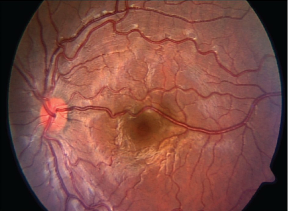
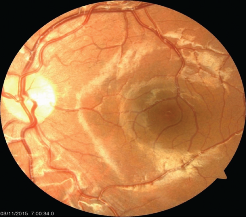
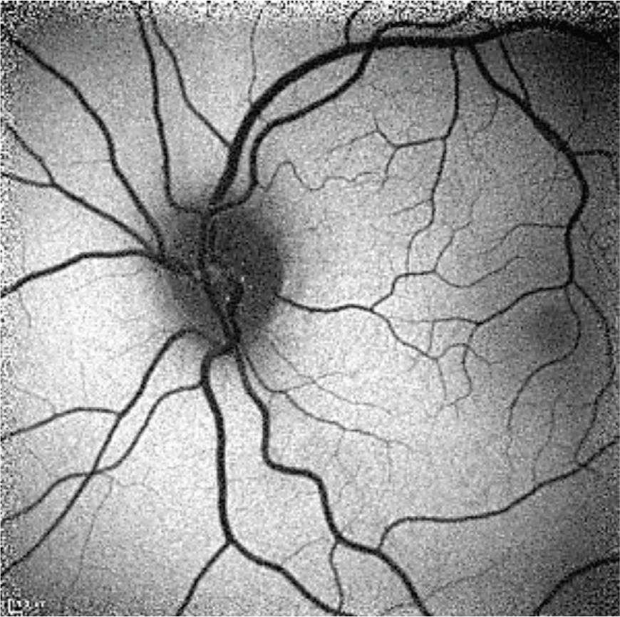
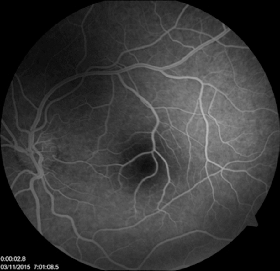
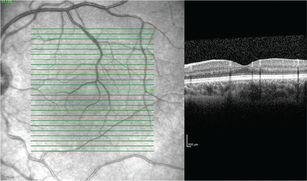
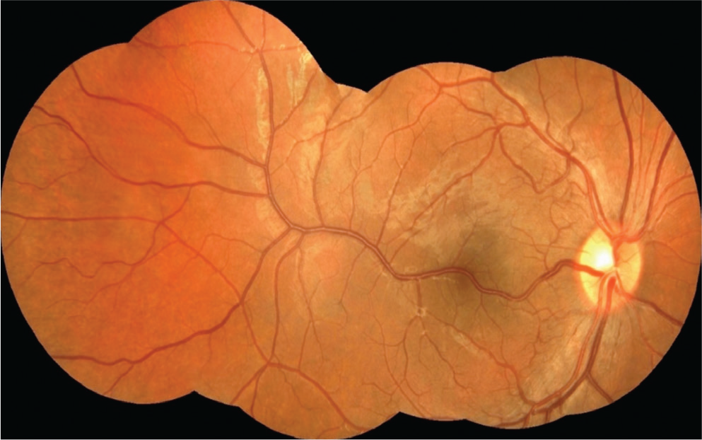
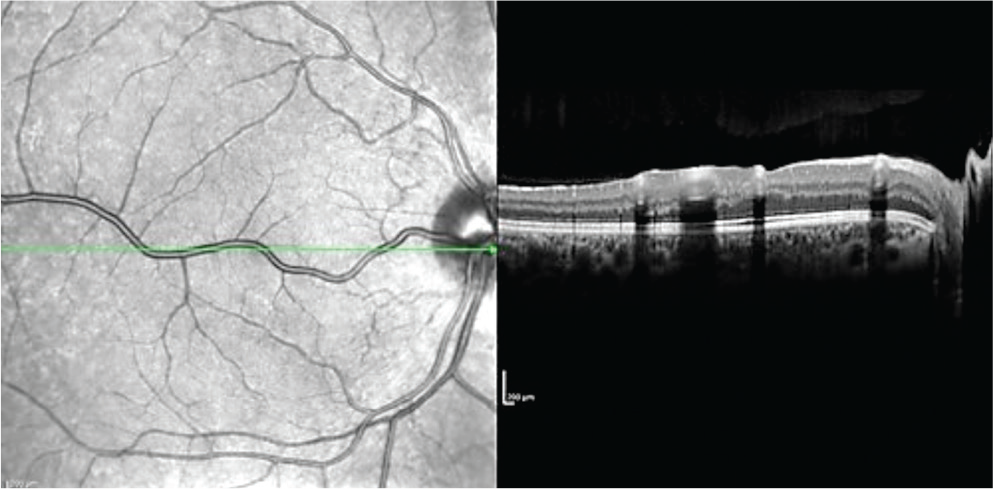
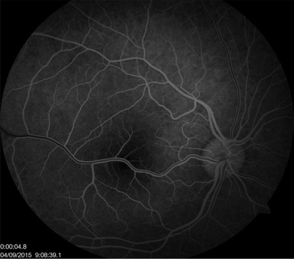
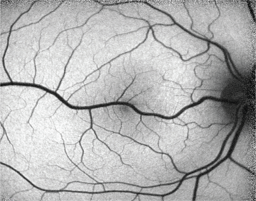
In this report, we describe three cases of retinal macrovessel. Two of the three patients presented to our clinic for annual eye exam and had no visual complaints. The third patient presented because of vision loss in the left eye. Two patients had 20/20 best corrected visual acuity in both eyes and the third patient had 20/20 in the right eye, 20/25 in the left eye. Pupillary exams were normal. Slit-lamp examinations of the anterior segment were unremarkable. Fundus examination revealed macrovessels in the left eyes of two patients and in the right eye of one patient. The patients underwent complete ophthalmological examinations including color fundus photography for all three patients and optic coherence tomography, fundus autoflorescence, and fundus fluorescein angiography for two of the patients. Cilioretinal artery coexisting with macrovessel was seen angiography in one case. Congenital retinal macrovessel is a rare vascular condition. It is often unilateral and the vessel is an aberrantly large branch of the retinal arteries or veins. They may cross the fovea and their visual impact is minimal. The coexistence of congenital retinal macrovessel and cilioretinal artery is very rare. Visual impairment may occur in congenital retinal macrovessel due to retinal cavernous hemangioma, foveal cysts, central serous retinopathy, and other retinal vascular abnormalities.
Keywords: Congenital retinal macrovessel, aberrant retinal vessels, cilioretinal artery
Introduction
Mauthner first reported a large aberrant retinal vessel crossing the macula in 1869. In 1982, Brown et al. described the clinical and fluorescein angiographic features of congenital retinal macrovessel (CRM) in seven patients. Impairment of vision in the involved eye is uncommon and is characterized by foveal cyst, macular hemorrhage, serous macular detachment, branch retinal artery occlusion or other vascular abnormalities. CRM occurs mainly in veins but more rarely may stem from an artery or artery and vein together. Beatty et al. in this case report we present a cilioretinal artery connecting with a CRM, suggesting that such patients are at increased risk of retinal vascular decompensation. Herein, we present three cases showing no vision loss in routine ophthalmological examination, and interestingly, one patient had both CRM and cilioretinal artery.
Discussion
Congenital retinal macrovessel is a rare finding and is usually discovered incidentally. CRM are mesenchymal in origin and develop around the first weeks of the second trimester when differentiation of arteries and veins occurs. They are generally asymptomatic, and vision is not affected in most cases. Archer et al. classified congenital retinal arteriovenous communications into three groups. Group 1 arteriovenous communications are the mildest variant, and clinically, can be very subtle. Group 2 are larger than those of group 1. Our case 2 was compatible with group 1 and our cases 1 and 3 were compatible with group 2 of the Archer classification. To our knowledge, a congenital retinal venous macrovessel that communicates with a cilioretinal artery is very rare. This condition was first described by Beatty et al. Most of the cases of CRM that have been documented to date exhibited normal visual acuity. When macrovessel is associated with reduced vision, one of the rare conditions should be considered: foveal cyst, macular hemorrhage or serous detachment, macular ischemia, branch retinal artery occlusion, and Valsalva retinopathy. For this reason, clinicians should be vigilant and follow these patients regularly.
References
Mauthner L. Lehrbuch der ophthalmoscopie. Tendler: Vienna; 1869. p. 249. [Google Scholar]
Brown GC, Donoso LA, Magargal LE, Goldberg RE, Sarin LK. Congenital retinal macrovessels. Arch Ophthalmol. 1982;100:1430–1436. [PubMed] [Google Scholar]
de Crecchio G, Alfieri MC, Cennamo G, Forte R. Congenital macular macrovessels. Graefes Arch Clin Exp Ophthalmol. 2006;244:1183–1187. [PubMed] [Google Scholar]
Sanfilippo CJ, Sarraf D. Congenıtal macrovessel associated wıth cystoid macular edema and an ipsilateral intracranial venous malformation. Retin Cases Brief Rep. 2015;9:357–359. [PMC free article] [PubMed] [Google Scholar]
Beatty S, Goodall K, adford R, Lavin MJ. Decompensation of a congenital retinal macrovessel with arteriovenous communications induced by repetitive rollercoaster rides. Am J Ophthalmol. 2000;130:527–528. [PubMed] [Google Scholar]
Bhatia HK, harma S, Laxminarayana P. Congenital Retinal Macrovessel with Normal Visual Acuity: A Case Report. Int J Ophthalmol Clin Res. 2015;2:2–4. [Google Scholar]
Archer DB, Deutman A, Ernest JT, Krill AE. Arteriovenous communications of the retina. Am J Ophthalmol. 1973;75:224–241. [PubMed] [Google Scholar]
Goel N, Kumar V, Seth A, Ghosh B. Branch retinal artery occlusion associated with congenital retinal macrovessel. Oman J Ophthalmol. 2014;7:96–97. [PMC free article] [PubMed] [Google Scholar]

