

Abstract
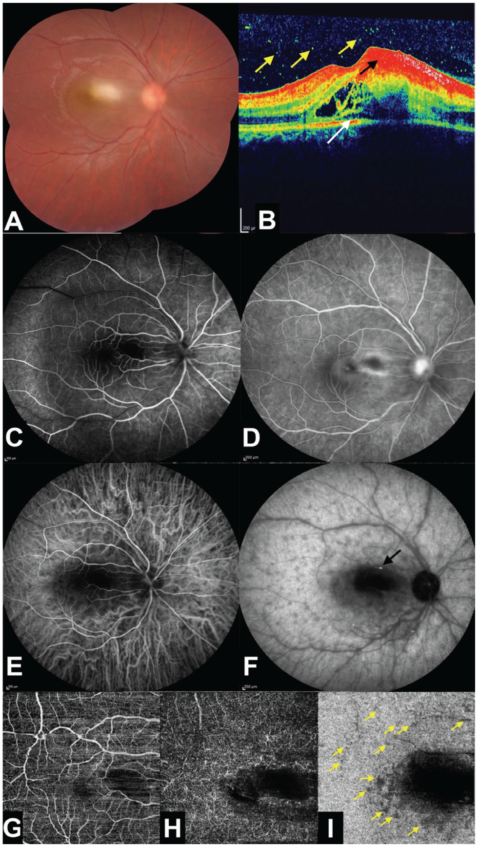
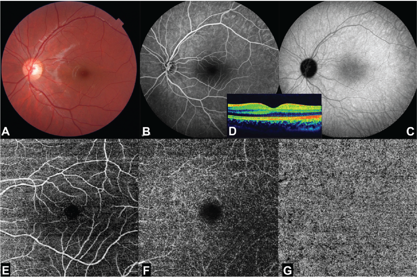
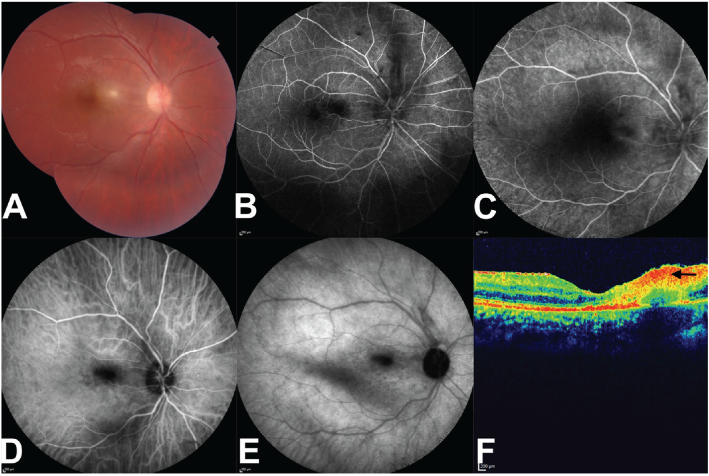
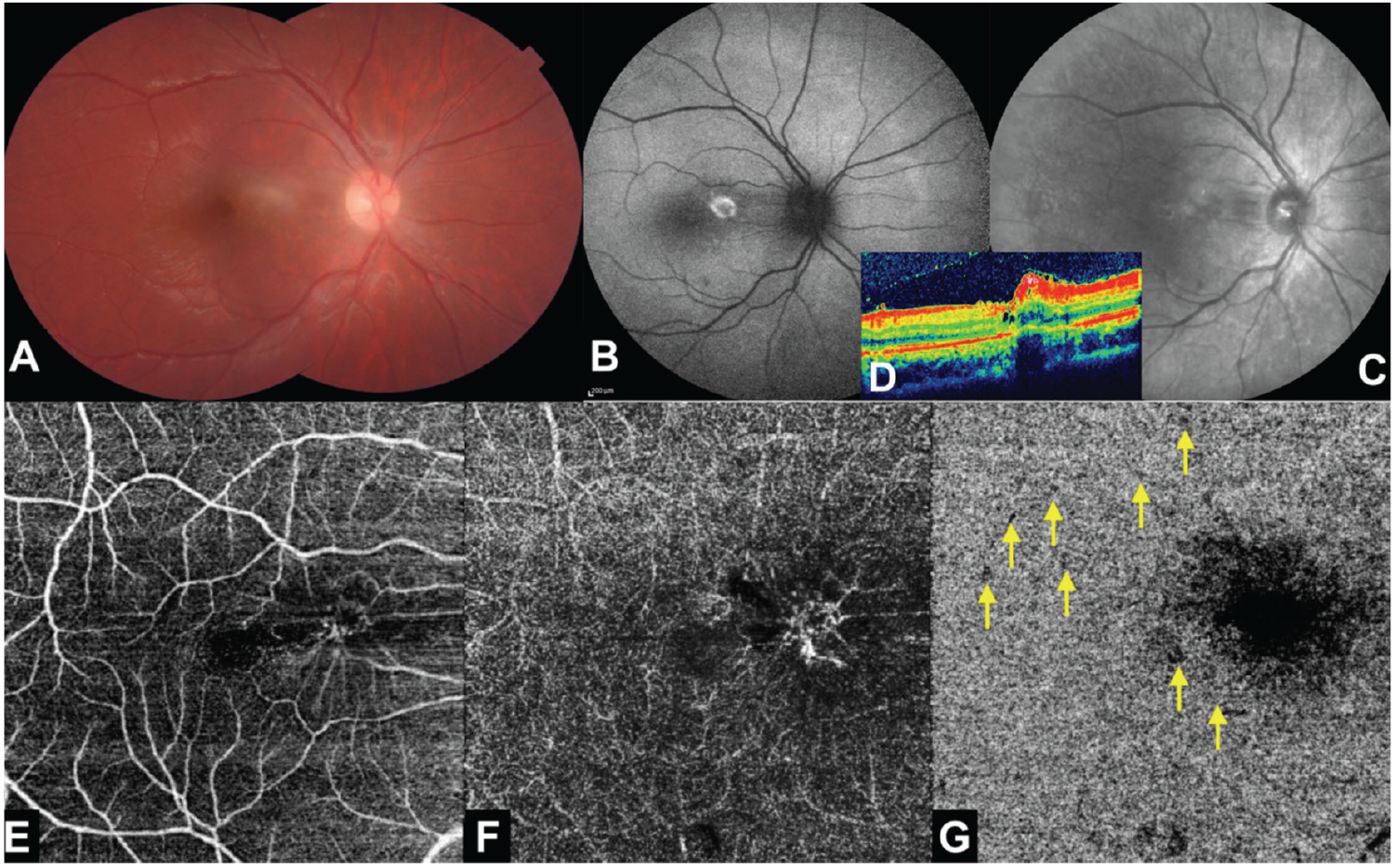
A 9-year-old otherwise healthy boy was examined due to a 4-day history of visual decline in his right eye. Ophthalmological examination revealed an area of active retinochoroiditis in the right macula. Indocyanine green angiography (ICGA) demonstrated multiple hypocyanescent dots surrounding the active lesion extending 360 degrees towards the equator. Optical coherence tomography angiography (OCTA) exhibited dark dots on the choriocapillaris slab over areas corresponding to the hypocyanescent dots detected with ICGA. Full systemic examination and laboratory investigations were carried out. Toxoplasma gondii serology was positive. The diagnosis of toxoplasmic chorioretinitis with white dot-like choroidal involvement was made. Trimethoprim/sulfamethoxazole, azithromycin, and oral prednisolone were administered orally. On repeated ICGA 2 weeks later, the scattered hypocyanescent dots were significantly fewer in number. A month later, right visual acuity was improved, the macular chorioretinitis focus had become inactive, an epiretinal membrane had formed, and the dark dots on the choriocapillaris slab of OCTA were markedly diminished. ICGA may be helpful to observe possible, subtle choroidal involvement in patients with toxoplasmic chorioretinitis.
Keywords: White dot syndrome, indocyanine green angiography, chorioretinitis, optical coherence tomography angiography, toxoplasmosis
Introduction
Toxoplasmic chorioretinitis is the most common type of infectious uveitis. Several atypical findings may accompany chorioretinitis such as scleritis, Fuchs’-like anterior uveitis, punctate outer retinitis, necrotizing retinitis, Coats’-type response, branch retinal artery occlusion, frosted branch angiitis-like retinal vasculitis, choroidal neovascular membrane, retinal detachment, papillitis, neuroretinitis, and retrobulbar neuritis. We herein report a 9-year-old boy with unilateral macular toxoplasmic chorioretinitis in association with multiple satellite dot-like choroidal involvement.
Discussion
Satellite dark dots in the choroid can be noted in eyes with active ocular toxoplasmosis.,, Knecht et al. argued that there was a secondary choriocapillaritis due to an inflammatory reaction of the choriocapillaris in the vicinity of the infection or inflammatory foci of the retina or choroid in many infectious uveitis entities including toxoplasmic chorioretinitis. They also speculated that satellite dark dots detected on ICGA might represent a temporary occlusion of the choriocapillaris likely related to a nonmechanical, cytokine-induced capillary occlusion.
Atmaca et al. retrospectively reviewed the charts and angiograms of 21 patients with ocular toxoplasmosis and noted hypofluorescent satellite dark dots adjacent to the main lesion in 11 of the eyes with active retinochoroiditis and they believed that hypersensitivity reaction might have played a role in the occurrence of these dots. However, the dark dots noted by Atmaca et al. did not present in a widespread white dot-like choroidal distribution as in our case. To our knowledge, 360-degree widespread white dot-like choroidal involvement observed in the present case has not been documented before, and we also shared the OCTA images of these dots.
In the present case, the 360-degree scattered satellite choroidal dots caught our attention only after viewing the sequence of ICGA, as those dots were very subtle both on color fundus photography and FA. Therefore, we believe that ICGA may be helpful to detect the presence of such satellite choroidal changes in patients with toxoplasmic chorioretinitis. These circumferentially scattered dark dots could be seen especially on the choriocapillaris slab of OCTA and they might either reflect impaired choroidal circulation or represent blockage due to inflammation-related coexistent subclinical inflammation or signs of a hypersensitivity reaction.
References
Auer C, Bernasconi O, Herbort CP. Indocyanine green angiography features in toxoplasmic retinochoroiditis. Retina. 1999;19:22-29.
Bernasconi O, Auer C, Herbor CP. Recurrent toxoplasmic retinochoroiditis Significance of perilesional satellite dark dots seen by indocyanine green angiography. Ocul Immunol Inflamm. 1997;5:207-211.
Auer C, Bernasconi O, Herbort CP. Toxoplasmic retinochoroiditis: New insights provided by indocyanine green angiography. Am J Ophthalmol. 1997;123:131-133.
Knecht PB, Papadia M, Herbort CP. Secondary choriocapillaritis in infectious chorioretinitis. Acta Ophthalmol. 2013;91:e550-e555.
Atmaca LS, Simsek T, Atmaca Sonmez P, Sonmez K. Fluorescein and indocyanine green angiography in ocular toxoplasmosis. Graefe’s Arch Clin Exp Ophthalmol. 2006;244:1688-1691.

