

Abstract
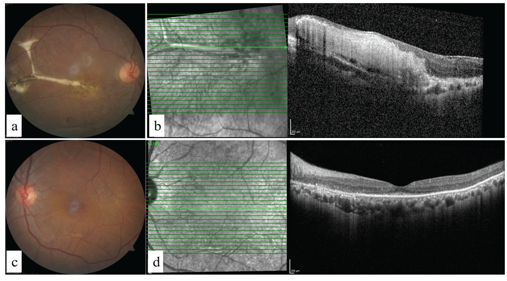
The aim of this case report is to describe a case of atypical central serous chorioretinopathy (CSCR) definitively diagnosed after 8 years. A 44-year-old woman presented with reduced visual acuity in her left eye. Her visual acuity was light perception with projection in the right eye and 0.15 in the left. She described a similar decline in vision in her right eye 8 years ago. At that time, she had exudative retinal detachment and was treated with systemic immunosuppressive therapy for a presumed diagnosis of Vogt-Koyanagi-Harada disease. Despite resolution of the exudative retinal detachment, macular scarring developed. Eight years later, she developed inferior exudative retinal detachment in the left eye. A diagnosis of atypical CSCR was made with the help of multimodal imaging and her left eye was successfully treated with eplerenone and half-fluence photodynamic therapy (hf-PDT). In conclusion, early diagnosis and treatment of atypical CSCR may prevent subretinal fibrosis formation and permanent vision loss. Hf-PDT and eplerenone are successful treatment options for atypical CSCR.
Keywords: Atypical central serous chorioretinopathy, indocyanine green angiography, photodynamic therapy, eplerenone
Introduction
Central serous chorioretinopathy (CSCR) is characterized by neurosensory macular detachment. However, in rare instances, the neurosensory detachment may be very extensive, causing bullous exudative retinal detachment. This rare variant of the disease is defined as atypical (bullous) CSCR. Characteristic fundoscopic features are multifocal exudative lesions in the posterior pole and inferior retinal detachment with shifting subretinal fluid.
Due to the unusual presentation, this form may be incorrectly diagnosed as rhegmatogenous retinal detachment, Harada disease, uveal effusion, multifocal choroiditis, metastatic carcinoma, or lymphoma. Inappropriate use of systemic corticosteroids and other immunosuppressive agents may cause exacerbation of the symptoms and lead to development of subretinal fibrosis and scarring.
Discussion
The pathogenesis of CSCR is not well documented. A breakdown in the permeability of the choriocapillaris has been implicated as the possible pathogenetic mechanism, leading to a focal loss of RPE-Bruch’s membrane attachment and allowing passage of the choroidal fluid into the subretinal space. Bullous retinal detachment is an extremely rare atypical variant of chronic CSCR which has been reported in a limited number of case presentations and case series.,
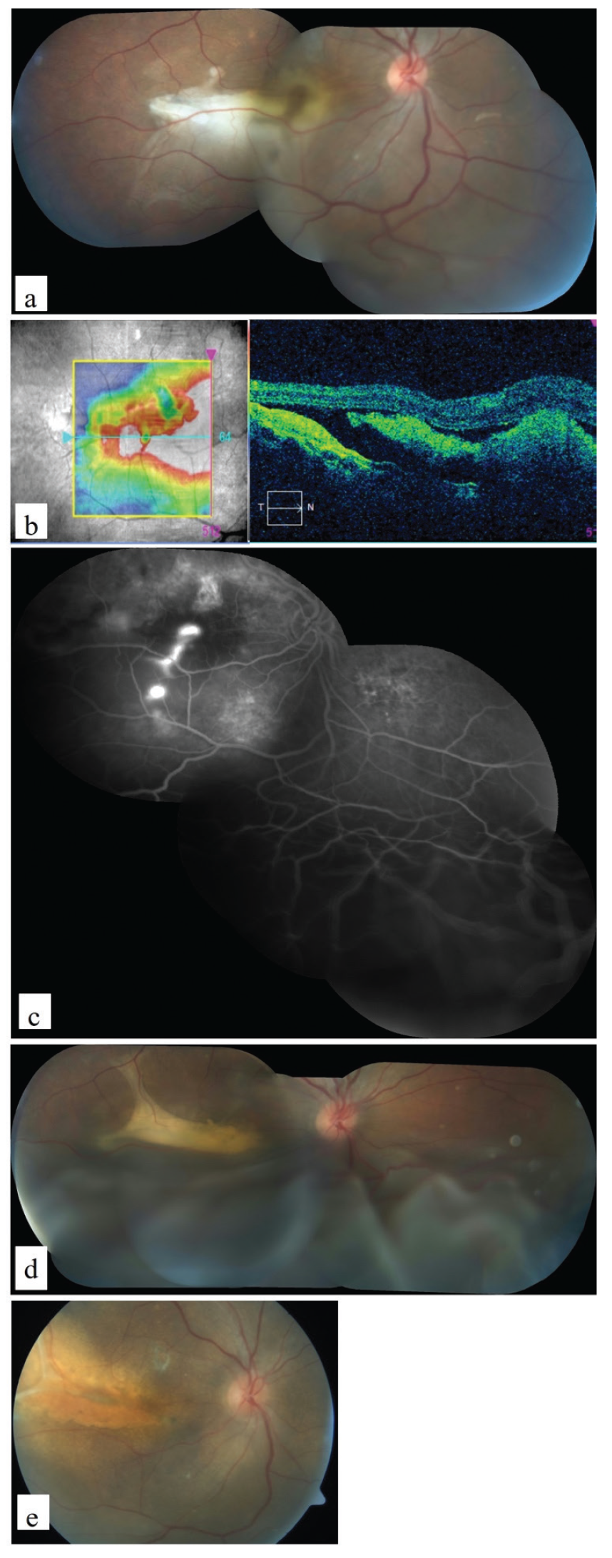
Corticosteroid therapy is one of the many systemic factors implicated in the pathogenesis of the bullous variant of CSCR. In our case, due to the presence of trace amount of vitreous cells, the patient was initially misdiagnosed with VKH disease and received intravenous high-dose corticosteroids. The use of steroids may aggravate the clinical findings of CSCR. Then, because the patient’s findings did not improve and her vision worsened, steroid-resistant VKH was suspected and her treatment was changed to other immunosuppressive and biologic agents.
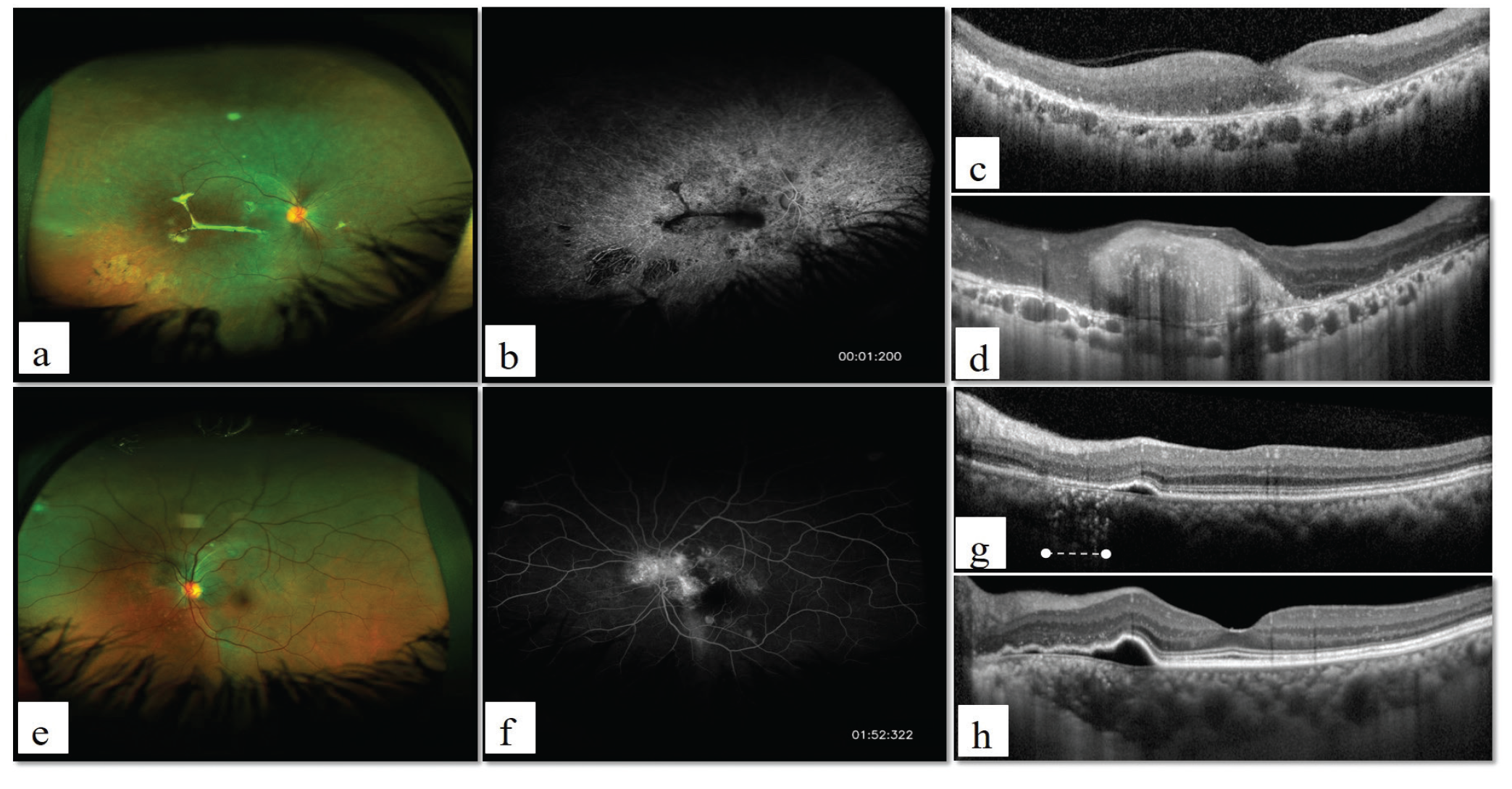
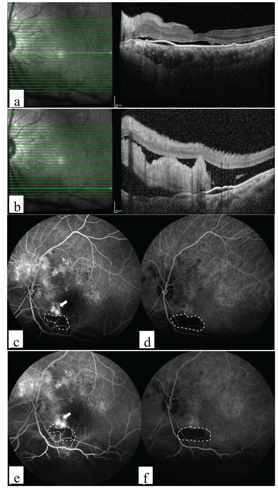
In atypical CSCR cases, providing an earlier definitive diagnosis may be a clinical challenge. Atypical bullous CSCR is most commonly misdiagnosed as the acute phase of VKH due to the exudative retinal detachment. Subretinal fibrin reaction and presence of generalized RPE irregularities in the absence of vitreous cells and lack of optic disc hyperemia and edema are signs in favor of CSCR. An absence of optic disc staining on FA and ICGA is a finding that further facilitates the diagnosis of CSCR. On OCT, RPE bulge may be seen in CSCR, whereas RPE folds, fluctuations of the internal limiting membrane, and subretinal septa are seen only VKH. Subretinal fibrin reaction is frequently encountered in eyes with bullous CSCR.
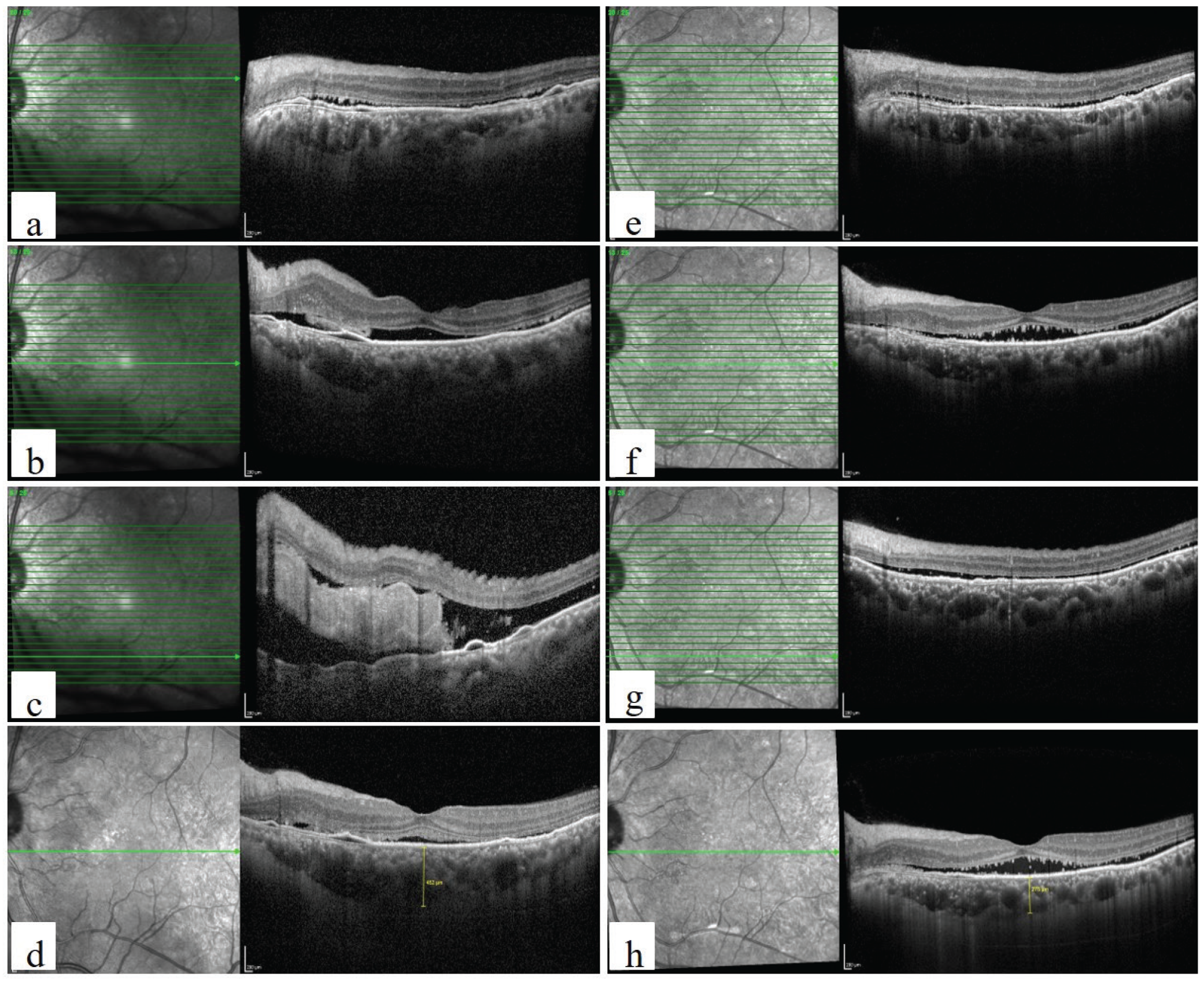
Therapeutic options for atypical CSCR include laser photocoagulation, PDT, and oral mineralocorticoid receptor antagonists. The main mechanism of action of PDT is angio-occlusion leading to constriction of choroidal vessels and choroidal vascular remodelling. Therefore, it may be the most appropriate treatment approach, being effective on the direct pathogenesis. In our case, we used a combination of half-fluence PDT and eplerenone therapy, which provided very rapid and complete resolution of subretinal fluid without complications.
In conclusion, the use of multimodal imaging may enable early definitive differential diagnosis of atypical CSCR from other chorioretinal diseases. Otherwise, inappropriate use of corticosteroids and other immunosuppressive agents may worsen the clinical findings and lead to poor visual prognosis. Hf-PDT in combination with oral eplerenone may be a successful treatment option for atypical CSCR that prevents subretinal fibrosis and scar formation.
References
Otsuka S, Ohba N and Nakao K. A long-term follow-up study of severe variant of central serous chorioretinopathy. Retina. 2002;22:25-32.
Hooymans JM. Fibrotic scar formation in central serous chorioretinopathy developed during systemic treatment with corticosteroids. Graefes Arch Clin Exp Ophthalmol. 1998;236:876-879.
Gass JDM. Stereoscopic Atlas of Macular Diseases: Diagnosis and Treatment. Mosby, 1997.
Gass JD and Little H. Bilateral bullous exudative retinal detachment complicating idiopathic central serous chorioretinopathy during systemic corticosteroid therapy. Ophthalmology. 1995;102:737-747.
Zmuda M, Tiev KP, Knoeri J and Heron E. Successful use of infliximab therapy in sight-threatening corticosteroid-resistant Vogt-Koyanagi-Harada disease. Ocul Immunol Inflamm. 2013;21:310-316.
Cebeci Z, Oray M, Bayraktar S, Tugal-Tutkun I and Kir N. Atypical Central Serous Chorioretinopathy. Turk J Ophthalmol. 2017;47:238-242.
Lin D, Chen W, Zhang G, Huang H, Zhou Z, Cen L, Chen H. Comparison of the optical coherence tomographic characters between acute Vogt-Koyanagi-Harada disease and acute central serous chorioretinopathy. BMC Ophthalmol. 2014;14:87.
Balaratnasingam C, Freund KB, Tan AM, Mrejen S, Hunyor AP, Keegan DJ, Dansingani KK, Dayani PN, Barbazetto IA, Sarraf D, Jampol LM, Yannuzzi LA. Bullous Variant of Central Serous Chorioretinopathy: Expansion of Phenotypic Features Using Multimethod Imaging. Ophthalmology. 2016;123:1541-1552.
Sartini F, Menchini M, Posarelli C, Casini G and Figus M. Bullous Central Serous Chorioretinopathy: A Rare and Atypical Form of Central Serous Chorioretinopathy. A Systematic Review. Pharmaceuticals (Basel) 2020;13.
Chan WM, Lam DS, Lai TY, Tam BS, Liu DT and Chan CK. Choroidal vascular remodelling in central serous chorioretinopathy after indocyanine green guided photodynamic therapy with verteporfin: a novel treatment at the primary disease level. Br J Ophthalmol. 2003;87:1453-1458.

