

Abstract
Pseudoexfoliation syndrome (PES) is one of the most common causes of open-angle glaucoma, with a higher risk of vision loss, a higher maximum and mean intraocular pressure (IOP) at diagnosis, and a wider range of IOP fluctuation compared to primary open-angle glaucoma. Patients with this syndrome have a ten-fold higher risk of developing glaucoma than the normal population. A definite diagnosis can be made by the observation of pseudoexfoliation material (PEM) on the anterior lens surface, ciliary processes, zonules, and iris. PEM deposits on the zonules may explain the clinically observed zonular weakness and lens subluxation or dislocation. An increased incidence of cataract development is also associated with PES. There is growing evidence for systemic associations of PES with peripheral, cardiovascular, and cerebrovascular system diseases, Alzheimer’s disease, hearing loss, and increased plasma homocysteine levels. Indications for surgery are markedly more common in patients with pseudoexfoliation glaucoma than primary open-angle glaucoma. The goal of this article is to review the latest perspectives on the clinical features, therapy, and systemic associations of this clinically and biologically challenging disease.
Keywords: Pseudoexfoliation, pseudoexfoliation syndrome, pseudoexfoliation glaucoma
Introduction
Pseudoexfoliation syndrome (PES) is the most common identified cause of open-angle glaucoma. Pseudoexfoliation is an independent risk factor for open-angle glaucoma in every country around the world. It is important to distinguish pseudoexfoliation glaucoma (PEG) from primary open-angle glaucoma (POAG). PEG is a severe type of glaucoma with a higher risk of vision loss, higher maximum and mean intraocular pressure (IOP) at diagnosis, and wider range of IOP fluctuation compared to POAG.
PEG may cause increased outflow resistance as a result of progressive accumulation of pseudoexfoliation material (PEM) in the trabecular meshwork, which contributes to alteration of retrobulbar blood flow and optic nerve microvascular blood flow, as well as elastosis of the lamina cribrosa.
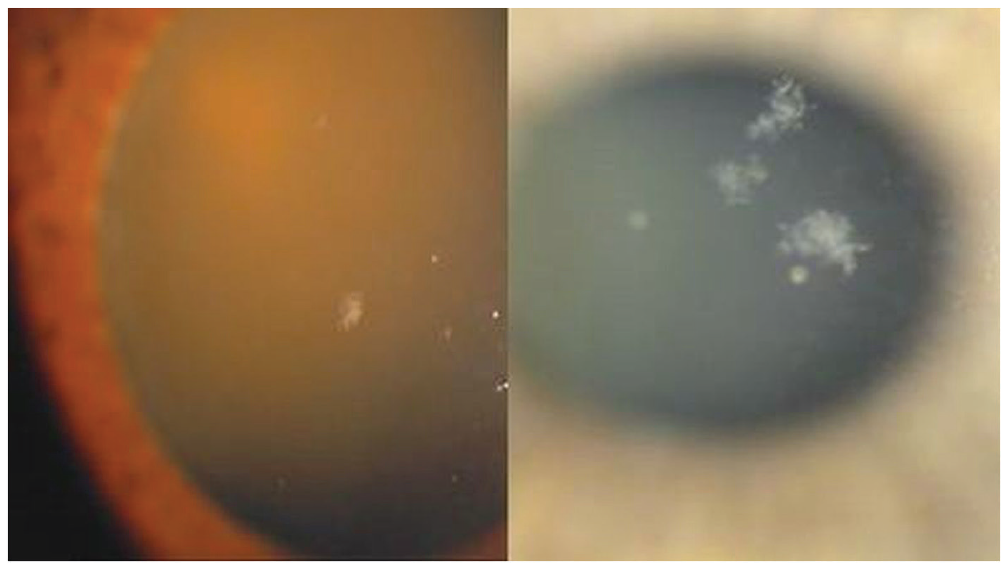
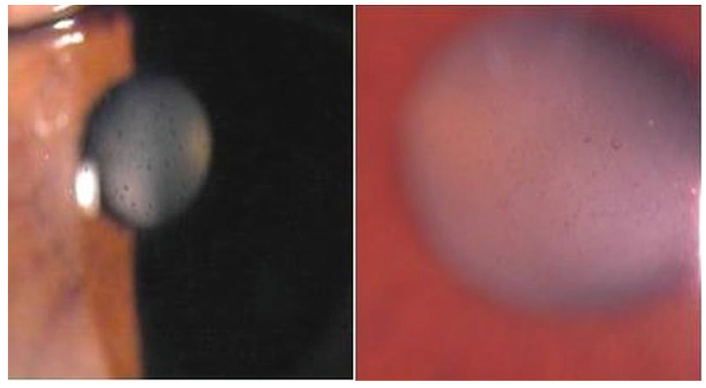
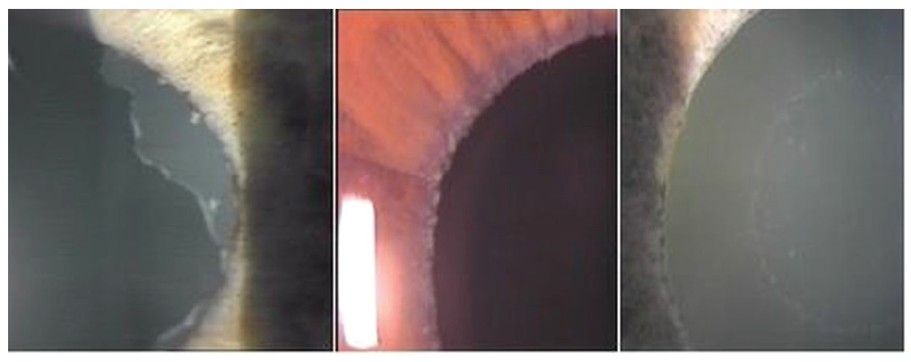
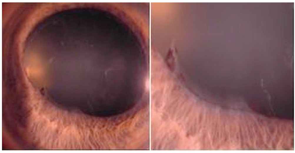
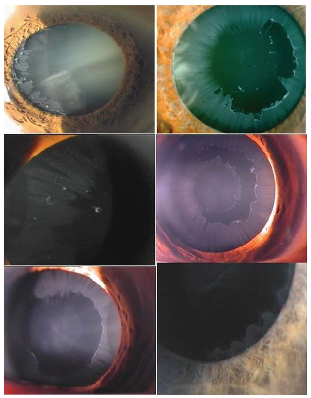
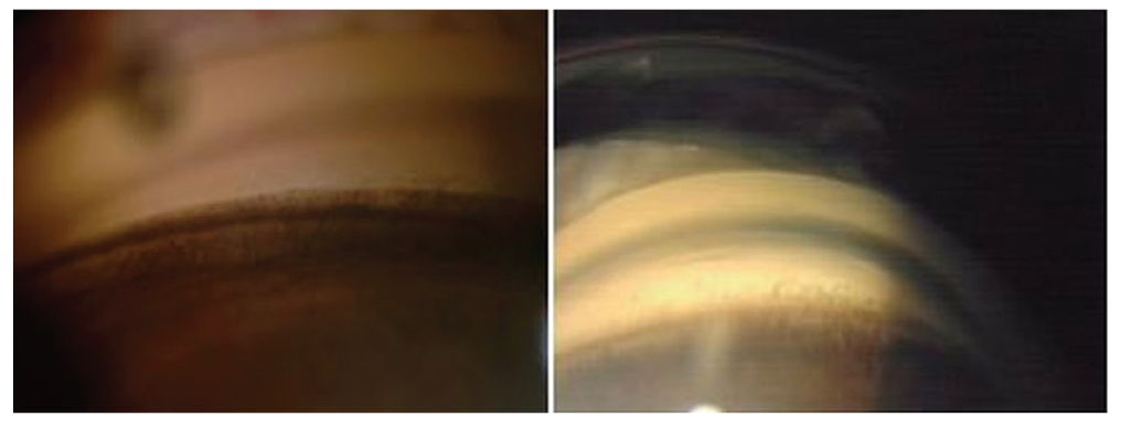
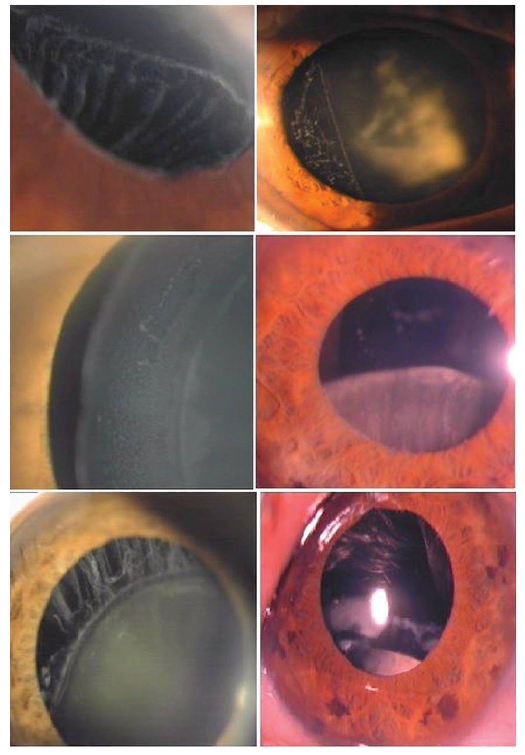
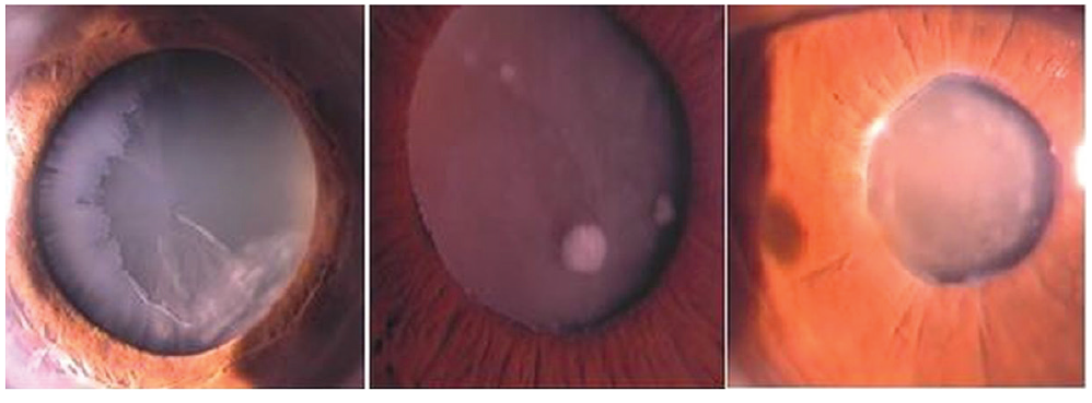
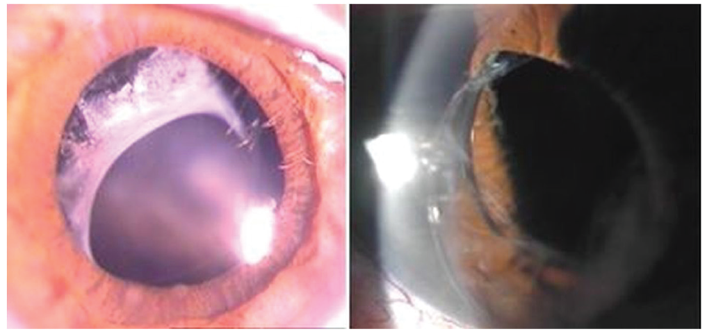
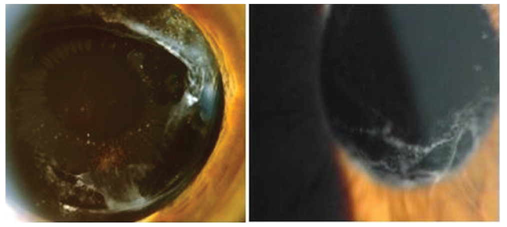
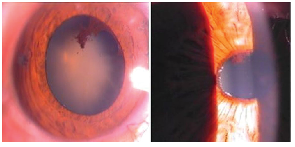
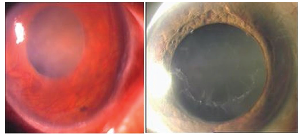
A definite diagnosis can be made by observing PEM on the anterior lens surface. These deposits can be found initially on the ciliary processes, zonules, and iris. Deposits of pseudoexfoliation on the zonules may explain the clinically observed zonular weakness and lens subluxation or dislocation. An increased incidence of cataract development is also associated with PES.
PES may be an important marker for cardiovascular and cerebrovascular diseases. There is growing evidence for systemic associations of PES with peripheral, cardiovascular, and cerebrovascular system diseases, Alzheimer’s disease, hearing loss, and increased plasma homocysteine levels.,
Clinical experiences show that controlling IOP is more difficult in PEG than in POAG. PEG patients with high IOP and advanced damage may benefit from a combination of anti-glaucoma medications as initial therapy. The indications for surgery are markedly more common in patients with PEG than in POAG.
The purpose of this article is to review current perspectives on the clinical features, therapy, and systemic associations of this clinically and biologically challenging disease.
Conclusions
PES and glaucoma are public health problems in older age worldwide. In recent years, the expansion in knowledge of the epidemiology, pathogenesis, and genetics of PES and glaucoma has provided important insights for understanding this disease and future treatment.
Pseudoexfoliation is an age-dependent and stress-induced extracellular fibrotic matrix disorder in which the oxidative-antioxidative balance is disturbed. LOXL1 is involved in the pathogenesis. Patients with PES have a higher risk of cardiovascular and cerebrovascular diseases.
The presence of pseudoexfoliation is an important risk factor for glaucoma and cataract. It is necessary to distinguish PEG from POAG. Medical management is more difficult and surgery is required more frequently. Early diagnosis, appropriate treatment and more frequent examinations appear to prevent the progression of visual field and vision loss.
In the future, new research can increase our understanding of the epidemiology, pathogenesis, genetics, and classifications of this disease. New therapeutic approaches will be considered for its management.
References
Ritch R, Schlötzer-Schrehardt U. Exfoliation syndrome. Surv Ophthalmol. 2001;45:265-315.
Mitchell P, Wang JJ, Smith W. Association of pseudoexfoliation syndrome with increased vascular risk. Am J Ophthalmol. 1997;124:685-687.
Altintas O, Yuksel N, Sonmez GT, Ozkan B, Altintas L, Caliskan Ş, Caglar Y. Serum antiphospholipid antibody levels in pseudoexfoliation. J Glaucoma. 2012;21:326-330.
Praveen MR, Shah SK, Vasavada AR, Diwan RP, Shah SM, Zumkhawala BR, Thomas R. Pseudoexfoliation as a risk factor for peripheral vascular disease: a case-control study. Eye (Lond). 2011;25:174-179.
Lindberg JG. Clinical investigations on depigmentation of the pupillary border and translucency of the iris in cases of senile cataract and in normal eyes in elderly persons. Acta Ophthalmol Suppl (1985). 1989;190:1-96.
Vogt A. Ein neues Spaltlampenbild des Pupillargebietes: Hellblauer Pupillarsaumfilz mit Hautchenbildung aus der Linsenvorderkapsel. Kin Monatsbl Atigenheilkd. 1925;75:1-12.
Dvorak-Theobald G. Pseudo-exfoliation of the lens capsule: relation to true exfoliation of the lens capsule as reported in the literature and role in the production of glaucoma capsulocuticulare. Am J Ophthalmol. 1954;37:1-12.
Cumurcu T, Kilic R, Yologlu S. The frequency of pseudoexfoliation syndrome in the middle Black Sea region of Turkey. Eur J Ophthalmol. 2020;20:1007-1011.
Forsius H. Exfoliation syndrome in various ethnic populations. Acta Ophthalmol Suppl (1985). 1988;184:71-85.
Ringvold A, Blika S, Elsås T, Guldahl J, Brevik T, Hesstvedt P, Johnsen H, Hoff K, Høisen H, Kjørsvik S, et al. The Middle-Norway eye-screening study. I. Epidemiology of the pseudo-exfoliation syndrome. Acta Ophthalmol (Copenh). 1988;66:652-658.
Anastasopoulos E, Topouzis F, Wilson MR, Harris A, Pappas T, Yu F, Koskosas A, Founti P, Coleman AL. Characteristics of pseudoexfoliation in the Thessaloniki Eye Study. J Glaucoma. 2011;20:160-166.
Mitchell P, Wang JJ, Hourihan F. The relationship between glaucoma and pseudoexfoliation: the Blue Mountains Eye Study. Arch Ophthalmol. 1999;117:1319-1324.
Ritch R. Exfoliation syndrome-the most common identifiable cause of open-angle glaucoma. J Glaucoma. 1994;3:176-177.
Vesti E, Kivelä T. Exfoliation syndrome and exfoliation glaucoma. Prog Retin Eye Res. 2000;19:345-368.
Koliakos GG, Konstas AG, Schlötzer-Schrehardt U, Bufidis T, Georgiadis N, Ringvold A. Ascorbic acid concentration is reduced in the aqueous humor of patients with exfoliation syndrome. Am J Ophthalmol. 2002;134:879-883.
Schlötzer-Schrehardt U. Molecular pathology of pseudoexfoliation syndrome/glaucoma--new insights from LOXL1 gene associations. Exp Eye Res. 2009;88:776-785.
Schlötzer-Schrehardt U. Genetics and genomics of pseudoexfoliation syndrome/glaucoma. Middle East Afr J Ophthalmol. 2011;18:30-36.
Thorleifsson G, Magnusson KP, Sulem P, Walters GB, Gudbjartsson DF, Stefansson H, Jonsson T, Jonasdottir A, Jonasdottir A, Stefansdottir G, Masson G, Hardarson GA, Petursson H, Arnarsson A, Motallebipour M, Wallerman O, Wadelius C, Gulcher JR, Thorsteinsdottir U, Kong A, Jonasson F, Stefansson K. Common sequence variants in the LOXL1 gene confer susceptibility to exfoliation glaucoma. Science. 2007;317:1397-1400.
Ritch R, Schlötzer-Schrehardt U. Exfoliation (pseudoexfoliation) syndrome: toward a new understanding. Proceedings of the First International Think Tank. Acta Ophthalmol Scand. 2001;79:213-217.
Schlötzer-Schrehardt U, Khor CC. Pseudoexfoliation syndrome and glaucoma: from genes to disease mechanisms. Curr Opin Ophthalmol. 2021;32:118-128.
Naumann GO, Schlötzer-Schrehardt U. Keratopathy in pseudoexfoliation syndrome as a cause of corneal endothelial decompensation: a clinicopathologic study. Ophthalmology. 2000;107:1111-1124.
Prince AM, Ritch R. Clinical signs of the pseudoexfoliation syndrome. Ophthalmology. 1986;93:803-807.
Schlötzer-Schrehardt UM, Koca MR, Naumann GO, Volkholz H. Pseudoexfoliation syndrome. Ocular manifestation of a systemic disorder?. Arch Ophthalmol. 1992;110:1752-1756.
Schlötzer-Schrehardt U, Naumann GO. Ocular and systemic pseudoexfoliation syndrome. Am J Ophthalmol. 2006;141:921-937.
Ritch R, Vessani RM, Tran HV, Ishikawa H, Tello C, Liebmann JM. Ultrasound biomicroscopic assessment of zonular appearance in exfoliation syndrome. Acta Ophthalmol Scand. 2007;85:495-499.
Guo S, Gewirtz M, Thaker R, Reed M. Characterizing pseudoexfoliation syndrome through the use of ultrasound biomicroscopy. J Cataract Refract Surg. 2006;32:614-617.
Sbeity Z, Dorairaj SK, Reddy S, Tello C, Liebmann JM, Ritch R. Ultrasound biomicroscopy of zonular anatomy in clinically unilateral exfoliation syndrome. Acta Ophthalmol. 2008;86:565-568
Zheng X, Sakai H, Goto T, Namiguchi K, Mizoue S, Shiraishi A, Sawaguchi S, Ohashi Y. Anterior segment optical coherence tomography analysis of clinically unilateral pseudoexfoliation syndrome: evidence of bilateral involvement and morphologic factors related to asymmetry. Invest Ophthalmol Vis Sci. 2011;52:5679-5684.
Altintas O, Yuksel N, Karabas VL, Caglar Y. Diurnal intraocular pressure variation in pseudoexfoliation syndrome. Eur J Ophthalmol. 2004;14:495-500.
Shihadeh WA, Ritch R, Scharf B, Liebmann JM. Delayed intraocular pressure elevation after pupillary dilation in exfoliation syndrome. Acta Ophthalmol. 2011;89:560-562.
Jeng SM, Karger RA, Hodge DO, Burke JP, Johnson DH, Good MS. The risk of glaucoma in pseudoexfoliation syndrome. J Glaucoma. 2007;16:117-121.
Topouzis F, Wilson MR, Harris A, Founti P, Yu F, Anastasopoulos E, Pappas T, Koskosas A, Salonikiou A, Coleman AL. Risk factors for primary open-angle glaucoma and pseudoexfoliative glaucoma in the Thessaloniki eye study. Am J Ophthalmol. 2011;152:219-228.e1.
Ritch R. Exfoliation syndrome. Curr Opin Ophthalmol. 2001;12:124-130.
Kozobolis VP, Christodoulakis EV, Naoumidi II, Siganos CS, Detorakis ET, Pallikaris LG. Study of conjunctival goblet cell morphology and tear film stability in pseudoexfoliation syndrome. Graefes Arch Clin Exp Ophthalmol. 2004;242:478-483.
Akdemir MO, Kirgiz A, Ayar O, Kaldirim H, Mert M, Cabuk KS, Taskapili M. The Effect of Pseudoexfoliation and Pseudoexfoliation Induced Dry Eye on Central Corneal Thickness. Curr Eye Res. 2016;41:305-310.
Öncel BA, Pinarci E, Akova YA. Tear osmolarity in unilateral pseudoexfoliation syndrome. Clin Exp Optom. 2012;95:506-509.
Sahin Atik S, Altın Ekin M. The role of Meibomian glands on the development of dry eye disease in patients with unilateral pseudoexfoliation. J Fr Ophtalmol. 2022;45:915-920.
Inoue K, Okugawa K, Oshika T, Amano S. Morphological study of corneal endothelium and corneal thickness in pseudoexfoliation syndrome. Jpn J Ophthalmol. 2003;47:235-239.
Naumann GO, Schlötzer-Schrehardt U, Küchle M. Pseudoexfoliation syndrome for the comprehensive ophthalmologist. Intraocular and systemic manifestations. Ophthalmology. 1998;105:951-968.
Wali UK, Bialasiewicz AA, Rizvi SG, Al-Belushi H. In vivo morphometry of corneal endothelial cells in pseudoexfoliation keratopathy with glaucoma and cataract. Ophthalmic Res. 2009;41:175-179.
Zheng X, Shiraishi A, Okuma S, Mizoue S, Goto T, Kawasaki S, Uno T, Miyoshi T, Ruggeri A, Ohashi Y. In vivo confocal microscopic evidence of keratopathy in patients with pseudoexfoliation syndrome. Invest Ophthalmol Vis Sci. 2011;52:1755-1761.
Shuba L, Nicolela MT, Rafuse PE. Correlation of capsular pseudoexfoliation material and iridocorneal angle pigment with the severity of pseudoexfoliation glaucoma. J Glaucoma. 2007;16:94-97.
Puska P, Tarkkanen A. Exfoliation syndrome as a risk factor for cataract development: five-year follow-up of lens opacities in exfoliation syndrome. J Cataract Refract Surg. 2001;27:1992-1998.
Sekeroglu MA, Bozkurt B, Irkec M, Ustunel S, Orhan M, Saracbasi O. Systemic associations and prevalence of exfoliation syndrome in patients scheduled for cataract surgery. Eur J Ophthalmol. 2008;18:551-555.
Drolsum L, Ringvold A, Nicolaissen B. Cataract and glaucoma surgery in pseudoexfoliation syndrome: a review. Acta Ophthalmol Scand. 2007;85:810-821.
Subasi S, Yuksel N, Karabas VL, Yilmaz Tugan B. Late in-the-bag spontaneous IOL dislocation: risk factors and surgical outcomes. Int J Ophthalmol. 2019;12:954-960.
Kristianslund O, Råen M, Østern AE, Drolsum L. Late in-the-bag intraocular lens dislocation: a randomized clinical trial comparing lens repositioning and lens exchange. Ophthalmology 2017;124:151-159.
Shingleton BJ, Nguyen BK, Eagan EF, Nagao K, O’Donoghue MW. Outcomes of phacoemulsification in fellow eyes of patients with unilateral pseudoexfoliation: single-surgeon series. J Cataract Refract Surg. 2008;34:274-279.
Akinci A, Batman C, Zilelioglu O. Phacoemulsification in pseudoex-foliation syndrome. Ophthalmologica. 2008;222:112-116.
Shingleton BJ, Crandall AS, Ahmed II. Pseudoexfoliation and the cataract surgeon: preoperative, intraoperative, and postoperative issues related to intraocular pressure, cataract, and intraocular lenses. J Cataract Refract Surg. 2009;35:1101-1120.
Hyams M, Mathalone N, Herskovitz M, Hod Y, Israeli D, Geyer O. Intraoperative complications of phacoemulsification in eyes with and without pseudoexfoliation. J Cataract Refract Surg. 2005;31:1002-1005.
Nagashima RJ. Decreased incidence of capsule complications and vitreous loss during phacoemulsification in eyes with pseudoexfoliation syndrome. J Cataract Refract Surg. 2004;30:127-131.
Bayraktar S, Altan T, Küçüksümer Y, Yilmaz OF. Capsular tension ring implantation after capsulorhexis in phacoemulsification of cataracts associated with pseudoexfoliation syndrome. Intraoperative complications and early postoperative findings. J Cataract Refract Surg. 2001;27:1620-1628.
Yüksel N, Doğu B, Karabaş VL, Cağlar Y. Foveal thickness after phacoemulsification in patients with pseudoexfoliation syndrome, pseudoexfoliation glaucoma, or primary open-angle glaucoma. J Cataract Refract Surg. 2008;34:1953-1957.
Gimbel HV, Condon GP, Kohnen T, Olson RJ, Halkiadakis I. Late in-the-bag intraocular lens dislocation: incidence, prevention, and management. J Cataract Refract Surg. 2005;31:2193-2204.
Pueringer SL, Hodge DO, Erie JC. Risk of late intraocular lens dislocation after cataract surgery, 1980-2009: a population-based study. Am J Ophthalmol. 2011;152:618-623.
Kozobolis VP, Glynatsis M, Labiris G, Katsanos A, Fanariotis M, Koukoula S, Alvanos S, Toufexis G. Retinal nerve fiber layer thickness in patients with exfoliation, exfoliative glaucoma, and primary open angle glaucoma. Eur J Ophthalmol. 2010;20:142-148.
Yüksel N, Altintaş O, Celik M, Ozkan B, Cağlar Y. Analysis of retinal nerve fiber layer thickness in patients with pseudoexfoliation syndrome using optical coherence tomography. Ophthalmologica. 2007;221:299-304.
Cursiefen C, Hammer T, Küchle M, Naumann GO, Schlötzer-Schrehardt U. Pseudoexfoliation syndrome in eyes with ischemic central retinal vein occlusion. A histopathologic and electron microscopic study. Acta Ophthalmol Scand. 2001;79:476-478.
Köse HC, Tekeli O. Optical coherence tomography angiography of the peripapillary region and macula in normal, primary open angle glaucoma, pseudoexfoliation glaucoma and ocular hypertension eyes. Int J Ophthalmol. 2020;13:744-754.
Subasi S, Yuksel N, Basaran E, Pirhan D. Comparison of vessel density in macular and peripapillary regions between primary open-angle glaucoma and pseudoexfoliation glaucoma using OCTA. Int Ophthalmol. 2021;41:173-184.
Cornelius A, Pilger D, Riechardt A, Reitemeyer E, Rübsam A, Winterhalter S, Maier AB. Macular, papillary and peripapillary perfusion densities measured with optical coherence tomography angiography in primary open angle glaucoma and pseudoexfoliation glaucoma. Graefes Arch Clin Exp Ophthalmol. 2022;260:957-965.
Lee JY, Sung KR, Kim YJ. Comparison of the Prevalence and Clinical Characteristics of Epiretinal Membrane in Pseudoexfoliation and Primary Open-angle Glaucoma. J Glaucoma. 2021;30:859-865.
Lee JY, Sung KR, Kim YJ. Association of Epiretinal Membrane With Pseudoexfoliation Glaucoma and Long-term Factors Affecting Visual Function. J Glaucoma. 2022;31:595-601.
Adıyeke SK, Kutlu N, Özen K, Doran MA, Demirbaş K, Ture G, Talay E. Is pseudoexfoliation syndrome associated with vitreoretinal interface abnormalities?. Graefes Arch Clin Exp Ophthalmol. 2022;260:431-437.
Streeten BW, Li ZY, Wallace RN, Eagle RC Jr, Keshgegian AA. Pseudoexfoliative fibrillopathy in visceral organs of a patient with pseudoexfoliation syndrome. Arch Ophthalmol. 1992;110:1757-1762.
Andrikopoulos GK, Mela EK, Georgakopoulos CD, Papadopoulos GE, Damelou AN, Alexopoulos DK, Gartaganis SP. Pseudoexfoliation syndrome prevalence in Greek patients with cataract and its association to glaucoma and coronary artery disease. Eye (Lond). 2009;23:442-447.
Demir N, Ulus T, Yucel OE, Kumral ET, Singar E, Tanboga HI. Assessment of myocardial ischaemia using tissue Doppler imaging in pseudoexfoliation syndrome. Eye (Lond). 2011;25:1177-1180.
Atalar PT, Atalar E, Kilic H, Abbasoglu OE, Ozer N, Aksöyek S, Ovünç K, Ozmen F, Gürsel E. Impaired systemic endothelial function in patients with pseudoexfoliation syndrome. Int Heart J. 2006;47:77-84.
Bourouki E, Oikonomou E, Moschos M, Siasos G, Siasou G, Gouliopoulos N, Deftereos S, Miliou A, Zacharia E, Tousoulis D. Pseudoexfoliative Glaucoma, Endothelial Dysfunction, and Arterial Stiffness: The Role of Circulating Apoptotic Endothelial Microparticles. J Glaucoma. 2019;28:749-755.
Repo LP, Suhonen MT, Teräsvirta ME, Koivisto KJ. Color Doppler imaging of the ophthalmic artery blood flow spectra of patients who have had a transient ischemic attack. Correlations with generalized iris transluminance and pseudoexfoliation syndrome. Ophthalmology. 1995;102:1199-1205.
Linnér E, Popovic V, Gottfries CG, Jonsson M, Sjögren M, Wallin A. The exfoliation syndrome in cognitive impairment of cerebrovascular or Alzheimer’s type. Acta Ophthalmol Scand. 2001;79:283-285.
Cumurcu T, Dorak F, Cumurcu BE, Erbay LG, Ozsoy E. Is there any relation between pseudoexfoliation syndrome and Alzheimer’s type dementia?. Semin Ophthalmol. 2013;28:224-229.
Aydoğan Ozkan B, Yüksel N, Keskin G, Altintaş O, Karabaş VL, Cağlar Y, Almaç A. Homocysteine levels in plasma and sensorineural hearing loss in patients with pseudoexfoliation syndrome. Eur J Ophthalmol. 2006;16:542-547.
Cahill M, Early A, Stack S, Blayney AW, Eustace P. Pseudoexfoliation and sensorineural hearing loss. Eye (Lond). 2002;16:261-266.
Singham NV, Zahari M, Peyman M, Prepageran N, Subrayan V. Association between Ocular Pseudoexfoliation and Sensorineural Hearing Loss. J Ophthalmol. 2014;2014:825936.
Yüksel N, Anik Y, Kiliç A, Karabaş V, Demirci A, Cağlar Y. Cerebrovascular blood flow velocities in pseudoexfoliation. Graefes Arch Clin Exp Ophthalmol. 2006;244:316-321.
Yüksel N, Anik Y, Altintaş O, Onur I, Cağlar Y, Demirci A. Magnetic resonance imaging of the brain in patients with pseudoexfoliation syndrome and glaucoma. Ophthalmologica. 2006;220:125-130.
Zikou AK, Kitsos G, Astrakas LG, Xydis VG, Spiliopoulos K, Bagli E, Argyropoulou MI. Pseudoexfoliation syndrome without glaucoma: White matter abnormalities detected by conventional MRI and diffusion tensor imaging. Eur J Radiol. 2018;99:82-87.
Altintaş O, Maral H, Yüksel N, Karabaş VL, Dillioğlugil MO, Cağlar Y. Homocysteine and nitric oxide levels in plasma of patients with pseudoexfoliation syndrome, pseudoexfoliation glaucoma, and primary open-angle glaucoma. Graefes Arch Clin Exp Ophthalmol. 2005;243:677-683.
Bleich S, Roedl J, Von Ahsen N, Schlötzer-Schrehardt U, Reulbach U, Beck G, Kruse FE, Naumann GO, Kornhuber J, Jünemann AG. Elevated homocysteine levels in aqueous humor of patients with pseudoexfoliation glaucoma. Am J Ophthalmol. 2004;138:162-164.
Roedl JB, Bleich S, Reulbach U, Rejdak R, Kornhuber J, Kruse FE, Schlötzer-Schrehardt U, Jünemann AG. Homocysteine in tear fluid of patients with pseudoexfoliation glaucoma. J Glaucoma. 2007;16:234-239.
Vessani RM, Ritch R, Liebmann JM, Jofe M. Plasma homocysteine is elevated in patients with exfoliation syndrome. Am J Ophthalmol. 2003;136:41-46.
Ritland JS, Egge K, Lydersen S, Juul R, Semb SO. Exfoliative glaucoma and primary open-angle glaucoma: associations with death causes and comorbidity. Acta Ophthalmol Scand. 2004;82:401-404.
Shrum KR, Hattenhauer MG, Hodge D. Cardiovascular and cerebrovascular mortality associated with ocular pseudoexfoliation. Am J Ophthalmol. 2000;129:83-86.
Kozart DM, Yanoff M. Intraocular pressure status in 100 consecutive patients with exfoliation syndrome. Ophthalmology. 1982;89:214-218.
Konstas AG, Mantziris DA, Stewart WC. Diurnal intraocular pressure in untreated exfoliation and primary open-angle glaucoma. Arch Ophthalmol. 1997;115:182-185.
Yüksel N, Karabaş VL, Arslan A, Demirci A, Cağlar Y. Ocular hemodynamics in pseudoexfoliation syndrome and pseudoexfoliation glaucoma. Ophthalmology. 2001;108:1043-1049.
Yüksel N, Karabaş VL, Demirci A, Arslan A, Altintaş O, Cağlar Y. Comparison of blood flow velocities of the extraocular vessels in patients with pseudoexfoliation or primary open-angle glaucoma. Ophthalmologica. 2001;215:424-429.
Netland PA, Ye H, Streeten BW, Hernandez MR. Elastosis of the lamina cribrosa in pseudoexfoliation syndrome with glaucoma. Ophthalmology. 1995;102:878-886.
Ritch R, Schlötzer-Schrehardt U, Konstas AG. Why is glaucoma associated with exfoliation syndrome?. Prog Retin Eye Res. 2003;22:253-275.
Ritch R. Perspective on exfoliation syndrome. J Glaucoma. 2001;10(5 Suppl 1):S33-S35.
Konstas AG, Hollo G, Astakhov YS, Teus MA, Akopov EL, Jenkins JN, Stewart WC. Factors associated with long-term progression or stability in exfoliation glaucoma. Arch Ophthalmol. 2004;122:29-33.
Konstas AG, Koliakos GG, Karabatsas CH, Liakos P, Schlötzer-Schrehardt U, Georgiadis N, Ritch R. Latanoprost therapy reduces the levels of TGF beta 1 and gelatinases in the aqueous humour of patients with exfoliative glaucoma. Exp Eye Res. 2006;82:319-322.
Konstas AG, Kozobolis VP, Katsimpris IE, Boboridis K, Koukoula S, Jenkins JN, Stewart WC. Efficacy and safety of latanoprost versus travoprost in exfoliative glaucoma patients. Ophthalmology. 2007;114:653-657.
Parmaksiz S, Yuksel N, Karabas VL, Ozkan B, Demirci G, Caglar Y. A comparison of travoprost, latanoprost, and the fixed combination of dorzolamide and timolol in patients with pseudoexfoliation glaucoma. Eur J Ophthalmol. 2006;16:73-80.
Yüksel N, Gök M, Altıntaş O, Cağlar Y. Diurnal intraocular pressure efficacy of the timolol-brimonidine fixed combination and the timolol-dorzolamide fixed combination as a first choice therapy in patients with pseudoexfoliation glaucoma. Curr Eye Res. 2011;36:804-808.
Konstas AG, Mikropoulos DG, Embeslidis TA, Dimopoulos AT, Papanastasiou A, Haidich AB, Stewart WC. 24-h Intraocular pressure control with evening-dosed travoprost/timolol, compared with latanoprost/timolol, fixed combinations in exfoliative glaucoma. Eye (Lond). 2010;24:1606-1613.
Konstas AG, Holló G, Mikropoulos D, Tsironi S, Haidich AB, Embeslidis T, Georgiadou I, Irkec M, Melamed S. Twenty-four-hour intraocular pressure control with bimatoprost and the bimatoprost/timolol fixed combination administered in the morning, or evening in exfoliative glaucoma. Br J Ophthalmol. 2010;94:209-213.
Goldenfeld M, Geyer O, Segev E, Kaplan-Messas A, Melamed S. Selective laser trabeculoplasty in uncontrolled pseudoexfoliation glaucoma. Ophthalmic Surg Lasers Imaging. 2011;42:390-393.
Gracner T. Intraocular pressure response of capsular glaucoma and primary open-angle glaucoma to selective Nd:YAG laser trabeculoplasty: a prospective, comparative clinical trial. Eur J Ophthalmol. 2002;12:287-292.
Konstas AG, Topouzis F, Leliopoulou O, Pappas T, Georgiadis N, Jenkins JN, Stewart WC. 24-hour intraocular pressure control with maximum medical therapy compared with surgery in patients with advanced open-angle glaucoma. Ophthalmology. 2006;113:761-5.e1.
Yilmaz Tugan B, Yuksel N, Kesim E, Subasi S. Comparison of long-term results of trabeculectomy and phacotrabeculectomy in patients with pseudoexfoliation glaucoma and primary open-angle glaucoma: a single-center study. Int Ophthalmol. 2022;42:1737-1747.
Drolsum L. Deep sclerectomy in patients with capsular glaucoma. Acta Ophthalmol Scand. 2003;81:567-572.
Drolsum L. Longterm follow-up after deep sclerectomy in patients with pseudoexfoliative glaucoma. Acta Ophthalmol Scand. 2006;84:502-506.
Rekonen P, Kannisto T, Puustjärvi T, Teräsvirta M, Uusitalo H. Deep sclerectomy for the treatment of exfoliation and primary open-angle glaucoma. Acta Ophthalmol Scand. 2006;84:507-511.
Subaşı S, Yüksel N, Özer F, Yılmaz Tugan B, Pirhan D. A Retrospective Analysis of Safety and Efficacy of XEN 45 Microstent Combined Cataract Surgery in Open-Angle Glaucoma over 24 Months. Turk J Ophthalmol. 2021;51:139-145.
Rauchegger T, Angermann R, Willeit P, Schmid E, Teuchner B. Two-year outcomes of minimally invasive XEN Gel Stent implantation in primary open-angle and pseudoexfoliation glaucoma. Acta Ophthalmol. 2021;99:369-375.
Sharkawi E, Lindegger DJ, Artes PH, Lehmann-Clarke L, El Wardani M, Misteli M, Pasquier J, Guarnieri A. Outcomes of gonioscopy-assisted transluminal trabeculotomy in pseudoexfoliative glaucoma: 24-month follow-up. Br J Ophthalmol. 2021;105:977-982.
Aktas Z, Ozdemir Zeydanli E, Uysal BS, Yigiter A. Outcomes of Prolene Gonioscopy Assisted Transluminal Trabeculotomy in Primary Open Angle Glaucoma and Pseudoexfoliation Glaucoma: A Comparative Study. J Glaucoma. 2022;31:751-756.

