

Dear Editor,
The PreserFlo® MicroShunt (PMS) implant (Santen, Miami, USA) is a newer alternative to traditional glaucoma surgeries like trabeculectomy. While the PMS is typically less effective at lowering intraocular pressure (IOP) compared to trabeculectomy, it is favored for its superior safety profile, including fewer reinterventions, and a lower risk of hypotony. However, the long-term success of PMS is influenced by fibrosis in the filtering bleb, which can increase IOP and lead to surgical failure. Postoperative needling procedures are often used to address bleb failure and restore bleb function. A rare and unique complication of PMS implantation is device dislocation, which can occur following needling procedures but has not been extensively documented in the medical literature. This article describes a case of PMS dislocation after an office-based needling procedure in a young patient.
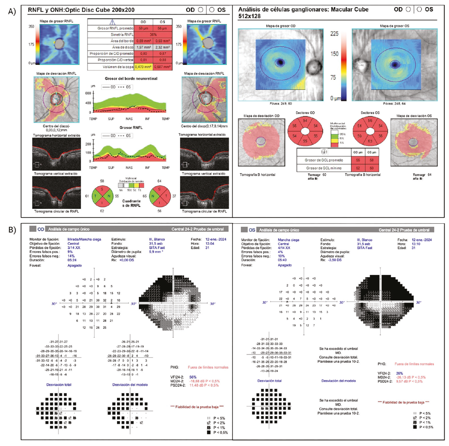
A 32-year-old man with a history of severe juvenile open-angle glaucoma in both eyes was monitored for ongoing IOP control. He had previously undergone multiple surgeries, including non-penetrating deep sclerectomy (NPDS) with revision and laser trabeculoplasty in the right eye, and NPDS, Ahmed valve implantation, cyclophotocoagulation, and revision in the left eye. Despite maximal topical therapy, IOP remained uncontrolled in the right eye. Preoperative examinations revealed visual acuity (Snellen decimal) of 0.7 in the right eye and 0.6 in the left eye, with IOPs of 40 mmHg and 15 mmHg, respectively. Both eyes showed signs of optic nerve damage, including significant pallor and thinning of the nerve fiber layer, as well as severe visual field involvement ().
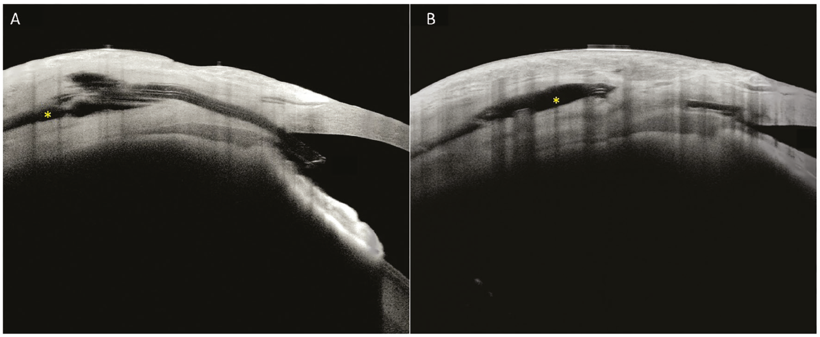
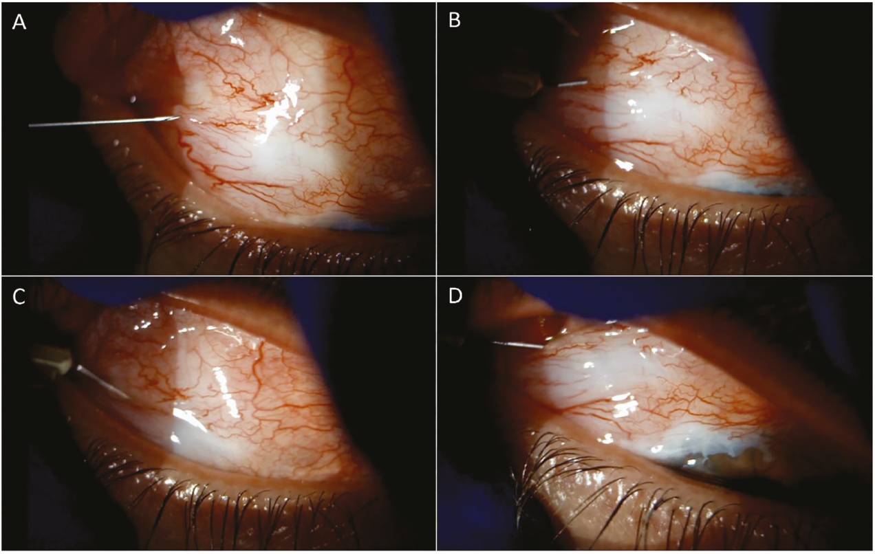
The patient had a prior history of hypotonic retinopathy after glaucoma surgery. Therefore, trabeculectomy was avoided, and ab externo PMS implantation was chosen for the right eye. The immediate postoperative course was uneventful, with an IOP of 9 mmHg and a well-formed bleb (). However, at 5 weeks, IOP increased to 18 mmHg, and anterior segment optical coherence tomography (AS-OCT) revealed contact between the distal end of the PMS and Tenon’s tissue (). A needling procedure was performed in the office to release the distal end (). The PMS was localized using a cannula and pressure on the conjunctiva to visualize the end of the implant. A 30-gauge (G) needle was used to make fan-shaped movements to release adhesions around the PMS. After the procedure, AS-OCT confirmed fluid around the PMS tip () and IOP dropped to 6 mmHg.
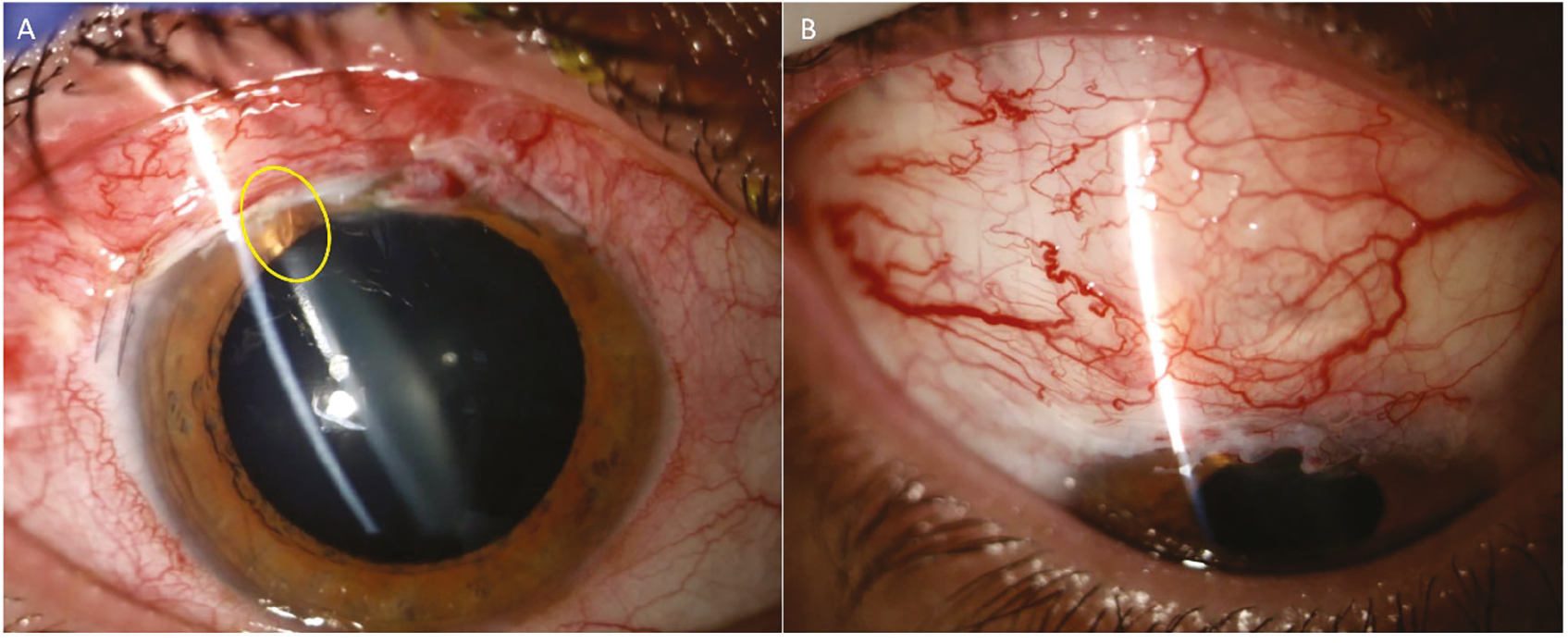
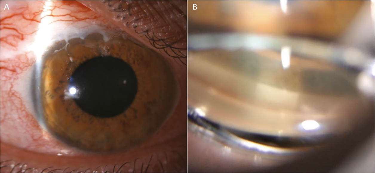
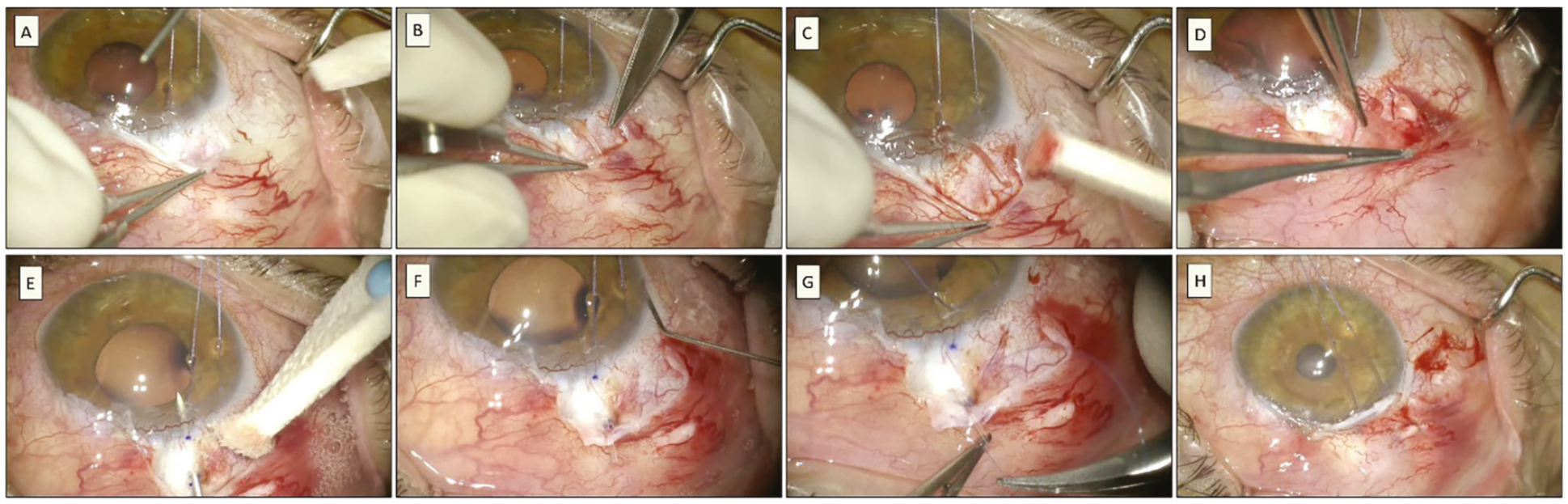
One month later, IOP remained below 14 mmHg, but 6 weeks post-needling, the patient presented with pain and a flattened bleb. IOP had risen to 50 mmHg, and the PMS was no longer visible in the anterior chamber (). Gonioscopy confirmed that the device was absent (). The patient was started on topical treatment and oral acetazolamide to reduce IOP, and a surgical revision was scheduled. During the revision surgery, the PMS was found to be deformed, rigid, and completely displaced posteriorly. The device had migrated out of the anterior chamber and was now located in the sub-Tenon’s space. The displaced PMS was removed, and a new PMS was implanted externally. Gonioscopy confirmed proper placement in the trabecular meshwork, and the device was guided with a 9-0 Prolene suture (). The immediate postoperative course was uneventful, and 2 months after the revision, IOP remained stable at around 16 mmHg, decreasing to 12 mmHg after ocular massage.
For several years, needling with or without subconjunctival antimetabolite injections has become a standard procedure to address bleb failure after trabeculectomy and PMS implantation. While needling is generally successful in restoring bleb function, it is not without risks. Although specific studies on complications after PMS needling are lacking, several case reports have described blood reflux, endophthalmitis, and device dislocation following needling with a 26G needle under the surgical microscope. This article presents the first reported case of PMS dislocation following an office-based needling procedure.
There are no established guidelines or widely described techniques for performing needling after PMS implantation. In this case, the surgeon used a 30G needle and fan-shaped movements above and below the distal end of the PMS to release adhesions obstructing aqueous humor flow. The procedure was conducted in the office under topical anesthesia, and the surgeon did not notice that the PMS was being displaced. As IOP decreased, the surgeon failed to recognize the changes in the anterior chamber visibility of the PMS. We hypothesize that the PMS remained in contact with the anterior chamber for a few days but eventually migrated out of the chamber due to blinking and other eye movements. Over the following weeks, fibrosis obstructed the drainage pathway, resulting in increased IOP and pain.
This case underscores the challenges of performing needling after PMS implantation. Unlike trabeculectomy, PMS implantation places the bleb in a smaller, more posterior area of the eye. This creates difficulty in freeing the distal end of the device from Tenon’s tissue while avoiding displacement during needle manipulation. Accurate localization of the end of the PMS is critical before performing needling. It is recommended to use a Hoskin lens or cannula and apply pressure to the conjunctiva to ensure proper visualization of the implant’s tip. Additionally, performing the procedure in an operating room setting provides more tools and better control if complications such as displacement occur.
Partial displacement of a PMS within the anterior chamber may not lead to a loss of therapeutic effect. However, if the device completely exits the anterior chamber, the surgical procedure will fail. The implant’s design allows for continued aqueous humor drainage until the drainage pathway becomes obstructed by fibrosis. This delayed failure makes diagnosis difficult, as the device may appear functional in the short term. When the PMS moves out of the anterior chamber, surgical revision is necessary.
In conclusion, minimally invasive glaucoma implants are increasingly important in glaucoma management. This case emphasizes that procedures to maintain bleb function carry potential complications. Special care during needling after PMS implantation is essential to prevent device dislocation, which can be difficult to diagnose and may ultimately result in surgical failure if the implant exits the anterior chamber.
Footnotes
Ethics
Informed Consent: Written informed consent was obtained from the patient for publication.
Authorship Contributions
Surgical and Medical Practices: L.S.V., M.C., Concept: R.G.R., Design: C.P.A., Data Collection or Processing: T.G.S., Analysis or Interpretation: M.T., Literature Search: P.G.V., P.G.B.E., Writing: R.G.R.
Conflict of Interest
Conflict of Interest: No conflict of interest was declared by the authors.
Financial Disclosure
Financial Disclosure: The authors declared that this study received no financial support.
References
Baker ND, Barnebey HS, Moster MR, Stiles MC, Vold SD, Khatana AK, Flowers BE, Grover DS, Strouthidis NG, Panarelli JF; INN005 Study Group. Ab-externo MicroShunt versus trabeculectomy in primary open-angle glaucoma: one-year results from a 2-year randomized, multicenter study. Ophthalmology. 2021;128:1710-1721.
Khan A, Khan AU. Comparing the safety and efficacy of Preserflo MicroShunt implantation and trabeculectomy for glaucoma: a systematic review and meta-analysis. Acta Ophthalmol. 2024;102:e443-e451.
Governatori L, Oliverio L, Mermoud A, Scampoli A, Sarati F, Carradori A, Catalani R, Monaco C, Caporossi T, Rizzo S. PreserFlo MicroShunt versus trabeculectomy: an updated meta-analysis and systematic review. Graefes Arch Clin Exp Ophthalmol. 2025;263:885-899.
Neubauer J, Suesskind D, Gassel CJ, Nasyrov E, Voykov B. Histopathological findings of failed blebs after microinvasive bleb surgery with the XEN Gel Stent and Preserflo MicroShunt. Graefes Arch Clin Exp Ophthalmol. 2024;262:2977-2984.
Hasan SM, Theilig T, Meller D. Comparison of Bleb Morphology following PRESERFLO® MicroShunt and trabeculectomy using anterior segment OCT. Diagnostics (Basel). 2023;13:3373.
Tanner A, Haddad F, Fajardo-Sanchez J, Nguyen E, Thong KX, Ah-Moye S, Perl N, Abu-Bakra M, Kulkarni A, Trikha S, Lascaratos G, Parnell M, Kailani O, King AJ, Agrawal P, Stead R, Giannouladis K, Rodrigues I, Goyal S, Hysi PG, Lim S, Yu-Wai-Man C. One-year surgical outcomes of the PreserFlo MicroShunt in glaucoma: a multicentre analysis. Br J Ophthalmol. 2023;107:1104-1111.
Cassottana P, Di Mola I, Ferro Desideri L, Vagge A, Cutolo CA, Traverso CE, Iester M. Blood reflux through a PreserFlo MicroShunt device after needling. J Glaucoma. 2022;31:979-981.
Brambati M, Bettin P, Ramoni A, Battista M, Bandello F. A case of endophthalmitis following needling procedure after PRESERFLO® Micro Shunt implantation. Eur J Ophthalmol. 2022;32:NP83-NP86.
Murakami K, Iida M, Shimada A, Ichioka S, Harano A, Tsutsui A, Tanito M. Dislocation of the PreserFlo MicroShunt during a postsurgical needling procedure. Cureus. 2023;15:e47356.

