

Öz
Hamartomlar normal olarak mevcut olduğu bölgede hücrelerin anormal çoğalmasından oluşan malformasyonlardır. Retina ve optik disk hamartomları başlığı altında astrositik hamartom, retina pigment epitelinin konjenital hipertrofisi (CHRPE), retina pigment epitelinin konjenital basit hamartomu (CSHRPE), retina ve retina pigment epitelinin kombine hamartomu (CHRRPE), retina hemanjiyoblastomu (retina kapiller hemanjiyomu) ve retina kavernöz hemanjiyomu yer alır. Retina ve optik disk hamartomları sistemik hastalıklar ile beraber olduğu gibi sporadik de izlenebilir. Astrositik hamartom genellikle düz sarı renkte ve şeffaf bir lezyondur. CHRPE yuvarlak, pigmentli, yüzeyden kabarıklık göstermeyen düz bir lezyondur. CSHRPE genellikle koyu siyah renkli, maküla yerleşimli bir tümör olarak ortaya çıkar. CHRRPE, vasküler, glial ve pigment epitel komponentlerinden oluşan, peripapiller, maküler ve periferik yerleşim gösterebilen bir kitledir. Retina hemanjiyoblastomu tipik olarak periferik retina veya optik diskte yerleşen kırmızı-pembe renkli, kıvrımlı ve dilate besleyici ve drene edici damarları olan vasküler bir tümördür. Retina kavernöz hemanjiyomu retinada veya optik sinir başında “üzüm salkımı benzeri” ince duvarlı sakküler anjiyomatöz yapıların oluşumu ile karakterizedir. Retina ve optik disk hamartomlarının tanısında ultrasonografi, fundus otofloresans, optik koherens tomografi, optik koherens tomogragi anjiyografi ve floresein anjiyografi yöntemleri kullanılmaktadır. Retina ve optik disk hamartomlarının bir kısmı tedavi gerektirmez. Ancak vitreus hemorajisi, maküler eksüdasyon, retina dekolmanı, maküler hol, epiretinal membran ve koroid neovaskülarizasyonu gibi komplikasyonlar gelişirse tedavi uygulanmalıdır.
Anahtar Kelimeler: Astrositik hamartom, retina pigment epitelinin konjenital hipertrofisi, retina pigment epitelinin konjenital basit hamartomu, retina ve retina pigment epitelinin kombine hamartomu, retina hemanjiyoblastomu, retina kavernöz hemanjiyomu
Giriş
Hamartom terimi Yunan dilinde hata anlamına gelen “hamartia” kelimesinden kaynaklanmaktadır. Hamartom normal olarak mevcut olduğu bölgede hücrelerin anormal çoğalmasından oluşan malformasyondur.1 Tek bir hücrenin mutasyona uğraması ile oluşan neoplaziden farklı olarak hamartomda birden fazla hücre etkilenir. Görünüm olarak normal dokuya benzeyen iyi huylu, yavaş büyüyen lezyonlardır ancak malign transformasyon meydana gelebilir. Hamartomlara genellikle genetik bir sendrom neden olur. Vücudun farklı bölgelerinde ortaya çıkabilir.1 Olguların çoğu asemptomatiktir ve diğer tıbbi durumların değerlendirilmesi sırasında tesadüfen tespit edilir.2
Retina ve optik disk hamartomları başlığı altında astrositik hamartom, retina pigment epitelinin konjenital hipertrofisi (CHRPE), retina pigment epitelinin konjenital basit hamartomu (CSHRPE), retina ve retina pigment epitelinin kombine hamartomu (CHRRPE), retina hemanjiyoblastomu (retina kapiller hemanjiyomu) ve retina kavernöz hemanjiyomu yer alır. Retina ve optik disk hamartomları sporadik veya sistemik hastalıklar ile beraber izlenebilir (). Astrositik hamartom, tüberoz skleroz kompleksi (TSK), nörofibromatozis tip 1, retinitis pigmentosa, Usher sendromu ve Stargardt hastalığında; CHRPE familyal adenomatöz polipozis (FAP) sendromunda; CHRRPE nörofibromatozis tip 1, nörofibromatozis tip 2, Gorlin-Goltz sendromu, Poland anomalisi ve brankiyookülofasiyal sendromda; retina hemanjioblastomu Von Hippel-Lindau (VHL) sendromunda; retina kavernöz hemanjiyomu serebral ve deri hemanjiyomlarını da içeren sendromik bir hastalık ile birlikte görülebilir.3,4,5,6,7
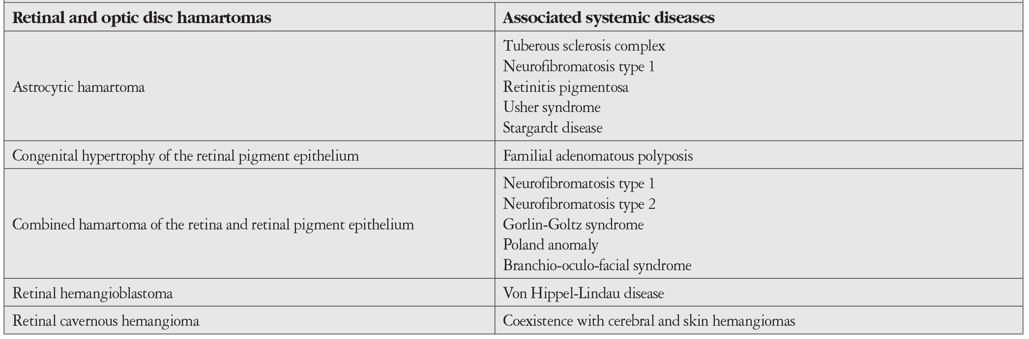
Literatürde retina ve optik disk hamartomlarını derleme şeklinde değerlendiren bir yayın mevcut değildir. Bu yazıdaki amacımız nadir görülen retina ve optik disk hamartomlarını topluca incelemektir.
Retina ve Optik Diskin Astrositik Hamartomu
Klinik Özellikler
Retina ve optik diskin astrositik hamartomu iyi huylu bir lezyondur. En sık TSK’li hastalarda görülür. İzole olarak veya diğer hastalıklara sekonder de izlenebilir. TSK’li olguların yaklaşık %50’sinde optik disk ve retina astrositik hamartomu mevcuttur.8 Astrositik hamartomu olan TSK’li olguların yaklaşık %30’unda bilateral tümörler ortaya çıkar.8 Astrositik hamartom daha nadiren nörofibromatozis tip 1, retinitis pigmentosa, Usher sendromu, Stargardt hastalığı ile birlikte görülebilir.8
Astrositik hamartom glial hücrelerden gelişir. Hamartin ve tuberini kodlayan TSC1 ve TSC2 genlerindeki mutasyonlar tümör gelişimine sebep olmaktadır.9,10 Retinada astrositik hamartom, fasial anjiyofibromlar ve deride vitiligoya benzeyen depigmente maküller TSK’li hastaların tipik triadıdır.8
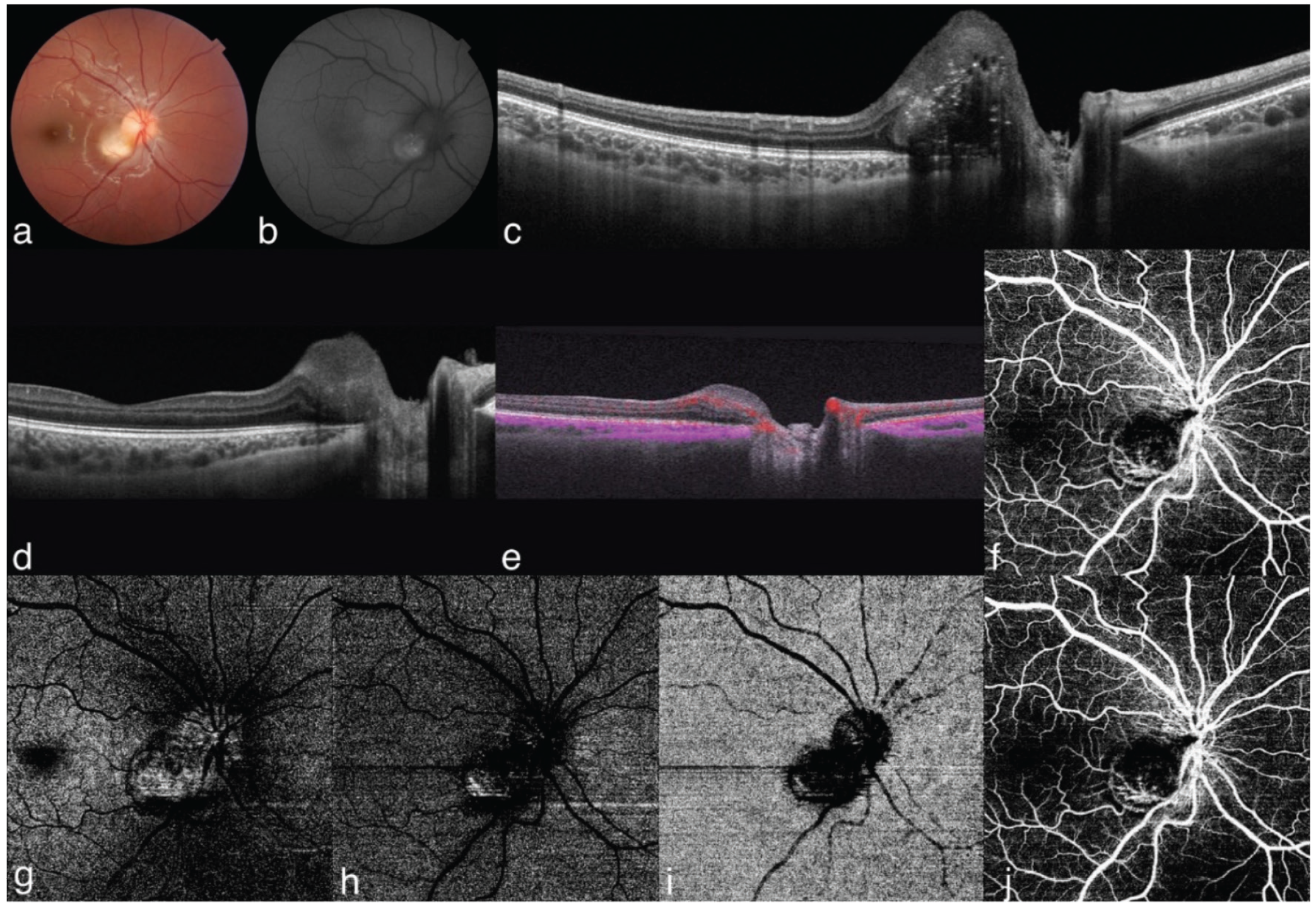
Astrositik hamartom genellikle düz, yuvarlak, şeffaf bir lezyon şeklinde ortaya çıkar (). Lezyon daha sonra büyüyerek nodüler bir yapı kazanır ve içinde kalsifikasyon gelişir.11,12 Bazen lezyonun ortası kalsifiktir; periferinde şeffaf kısım bulunabilir. Astrositik hamartom çevresinde sert eksüdaya rastlanabilir.13 Sert eksüda, tedavisiz retinoblastom/retinositomda genellikle görülmez.13,14 Buna karşılık, retinoblastom ve retinositomlarda izlenen retina pigment epiteli (RPE) değişiklikleri de astrositik hamartomlarda genellikle ortaya çıkmaz.14 Astrositik hamartomdaki kalsifikasyon parlak sarı renkte ve balık yumurtalarına benzer görünümdedir.15 Halbuki retinoblastomdaki kalsifikasyon tebeşir beyazı rengindedir.15 Ayırıcı tanısında düşünülmesi gereken diğer hastalıklar arasında optik disk ödemine neden olan durumlar, optik sinir başı druzeni, akkiz retinal astrositomlar ve reaktif gliozis yer alır.
Astrositik hamartom genelde stabil seyir gösterse de dev hücreli astrositomlarda progresif büyüme, sekonder glokom ve bulbus yıkımı görülebilir.15 Bu tümörler malign olarak kabul edilir ama metastaz yapmaz.15
İnceleme Yöntemleri
Astrositik hamartom ultrasonografik incelemede akustik solid kitle olarak görülür ve lezyon içinde kalsifikasyon izlenir. Astrositik hamartom fundus otofloresans (FAF) görüntülemesinde kalsiyum içeriğine bağlı olarak hiperotofloresans gösterir ().16 Optik koherens tomografide (OKT) kubbe veya plato şeklinde, orta-yüksek reflektivite gösteren ve koroid gölgelenmesi yapan lezyondur ().16 Koryokapillaris korunmuştur ve güve-yeniği boşlukları izlenebilir. OKT-anjiyografide (OKTA) iyi sınırlı, hiperreflektif lezyon ve ince dallanan tümör damarları izlenir.16,17 B-mod anjiyografide tümör içinde retinal akım sinyalleri tespit edilebilir (). Yüzeysel ve derin retinada tümör damar ağı görülür (). Dış retina ve koryokapillaris, lezyon içindeki kalsiyumdan veya yüksek kan akışından kaynaklanan gölgelenme/maskeleme nedeniyle hiporeflektif değişiklikler gösterir (Şekil 1h,i). Lezyon içindeki güve yeniği boşluklarına bağlı derin retinal pleksusta akım olmayan alanlar görülür ve dış retina ve koryokapillariste projeksiyon artefaktları izlenebilir.16 Tüm maküla kompozit görüntüde lezyon hiporeflektif olarak görülür (Şekil 1j). Lezyon santralinde derin kapiller pleksusta izlenen tümör vasküler yapısına ait hiperreflektif sinyaller alınmaktadır. Floresein anjiyografide (FA) koroidal fazda erken blokaj görülebilir, arter safhasında entrensek tümör damarlarında dolum başlar ve hiperfloresans giderek artar. Geç venöz fazda sızıntı olabilir. Astrositik hamartomun ayırıcı tanısında retinoblastom, retinositom, myelinli sinir lifleri, masif retinal gliozis gibi tanılar düşünülebilir.11,12,18
Klinik ve OKT bulgularına göre retina astrositik hamartomunun dört tipi belirlenmiştir (Pichi sınıflandırması).19 Tip 1’de OKT’de düz retina lezyonu mevcuttur (<500 µm) ve klinik olarak retina traksiyonu görülmez. Tip 2’de OKT’de hafif kabarık retina lezyonu izlenir (>500 µm) ve klinik olarak retina traksiyonu mevcuttur. Tip 3’te OKT’de iç retinada kalsifikasyon gösteren kabarık lezyon görülür (>500 µm) ve klinik olarak “dut benzeri” kalsifikasyon izlenir. Tip 4’te OKT’de optik boş kaviteleri olan kabarık lezyon görülür ve klinik olarak iç retinada düz non-kalsifiye lezyon mevcuttur. Tip 2 retina astrositik hamartomlar fibröz deri plakları, Tip 3 subependimal dev hücreli astrositik hamartomu ve Tip 4 akciğer lenfanjiyomiyomatozisi olan TSK olgularında istatistiksel anlamlı olarak daha fazla görülmektedir.19
Tedavi ve Prognoz
Astrositik hamartom genellikle stabil seyreder ve tedaviye gerek olmaz. Nadiren spontan regresyon gelişebilir.20 Vitreus hemoraji, retinal eksüdasyon, retina dekolmanı ve neovasküler glokom gelişme riskinden dolayı periyodik izlem önemlidir. Küçük lezyonlarda laser fotokoagülasyon uygulanabilir. Daha büyük ve semptomatik (eksüda, sıvı varsa) tümörlerde fotodinamik tedavi denenebilir. Sekonder koroid neovaskülarizasyonu mevcutsa anti vasküler endotelyal büyüme faktör (anti-VEGF) enjeksiyonu yapılabilir. Tümör içi ince damarlardan veya neovaskülarizasyondan kaynaklanan vitreus hemorajisi gelişmişse pars plana vitrektomi cerrahisi gerekebilir.
Dev hücreli astrositik hamartom agresif bir seyir gösterebilir; tümörde nekroz, vitreus hemorajisi, subretinal hemoraji, masif eksudasyon ve neovasküler glokom gelişmesi sonrası eksüdasyon gerekebilir.21 Neovasküler glokom, tümör içi nekroz veya kronik retina dekolmanı sonucu ortaya çıkmaktadır. Subependimal dev hücreli astrositom ve renal anjiyomiyolipom mevcut olan TSK olgularında m-TOR inhibitörleri (rapamycin, everolimus, sirolimus) kullanılabilir.22
Retina Pigment Epitelinin Konjenital Hipertrofisi (CHRPE)
Klinik Özellikler
Yuvarlak, pigmentli, yüzeyden kabarıklık göstermeyen düz bir fundus lezyonudur. RPE seviyesinde yerleşimlidir. Soliter, multifokal ve atipik olmak üzere üç varyantı mevcuttur (). CHRPE lezyonları, histopatolojik olarak, aşırı pigment granülleri içeren hipertrofik RPE hücrelerinden ibarettir. Koryokapillaris ve koroid normaldir. RPE üstündeki fotoreseptör tabakası normal veya atrofik olabilir. Lezyon kronikleştikçe fotoreseptör atrofisi tam olarak ortaya çıkar.23 Fotoreseptör atrofisi gelişen olguların görme alanı incelemesinde absolü skotomlar izlenir.24
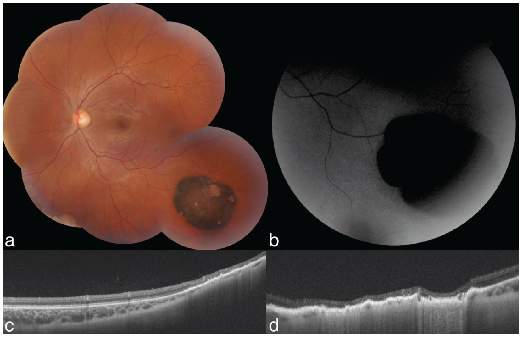
CHRPE genellikle düzgün kenarlı bir lezyondur; nadiren düzensiz kenarlı olabilir. Sıklıkla periferik fundusda ortaya çıkar. Peripapiller yerleşim daha nadirdir. Lezyon içinde depigmente lakünalar ve etrafında hipopigmente halo bulunabilir (). CHRPE lezyonları özellikle miyopik gözlerde minimal büyüme gösterebilirler.23 CHRPE’den nadiren RPE adenomu/adenokarsinomu tarzında lezyonlar gelişebilir.25
CHRPE nadiren amelanotik bir lezyon olabilir.26 Bu tip lezyonlara amelanotik CHRPE denir. Lezyon üzerindeki retina ve retina damarları normaldir.26 Fokal intraretinal pigmentasyon bulunabilir. Nadiren CHRPE kenarında neovaskülarizasyon görülebilir.27
CHRPE’nin bir tipi FAP ile birlikte ortaya çıkar. FAP genine APC (adenomatöz polipozis coli) geni denir ve 5q21-q22 bölgesinde lokalizedir. FAP ile birlikte bulunan CHRPE, genellikle oval şekillidir ve kuyrukları vardır.28 Bu lezyonlar, CHRPE’den ayırt edilebilmek için pigmentli oküler fundus lezyonları (pigmented ocular fundus lesions- POFL) olarak adlandırılmıştır.29 Bu lezyonların kuyrukları depigmente olabilir. İçlerinde lakünalar bulunabilir. Histopatolojik olarak CHRPE’ye benzer şekilde RPE hipertrofisi, hiperplazisi ve hamartomu tarzında patolojik görünümleri mevcuttur.29 Pigmentli oküler fundus lezyonlarının taban çapı genellikle 5 mm’den küçüktür.30
Pigmentli oküler fundus lezyonları, Gardner sendromu ve Turcot sendromu ile beraber görülebilir.29 Gardner sendromunda kolonda yüzlerce polip ortaya çıkar. Bu poliplerden kolon adenokarsinomu meydana gelir. Profilaktik kolektomi önerilir. Gardner sendromunda görülen ekstrakolonik kanserler tiroid, adrenal bez ve karaciğerde görülür. İyi huylu lezyonlar kafa ve orbitada gelişen osteom, sebase kist, lipom ve fibromlardır. Opak çene lezyonları da izlenebilir. Genellikle 4 veya daha fazla POFL görülmesi FAP yönünde önemli bir bulgudur.29 Turcot sendromunda ise POFL görülmesi yanında hastalarda beyin tümörü görülür.31
İnceleme Yöntemleri
Ultrasonografik incelemede CHRPE lezyonları düz veya minimal kabarık (<0,5 mm), akustik solid lezyonlar olarak görülür. FAF görüntülemesinde lezyonlarda yüksek melanin içeriğinden dolayı tipik olarak hipootofloresans görülür ().16 Pigmentli olmayan halolar veya boşluklar otofloresans gösterebilir.32 OKT’de düz, RPE düzeyinde yüksek reflektif birikimler gösteren ve koroid gölgelenmesi yapan lezyonlardır ().16 İç retina katları normaldir ama dış retina katlarında incelme saptanır.16 OKTA’da CHRPE genelde iyi sınırlı, yüzeysel ve derin retinal pleksusda hiperreflektif görünümdedir.16 B-mod anjiyografide tümör üzerindeki retinada akış sinyalleri izlenebilir.16 Dış retina ve koryokapillarisin maskelenmesi, “signal void” alanların görülmesine neden olmaktadır.16 FA’da sızıntı görülmez.24 Depigmente halolar veya boşlukların olduğu alanlar dışındaki bölgelerde alttaki koroid flöresansının blokajı izlenir.24
Tedavi ve Prognoz
CHRPE lezyonları genelde tedavi gerektirmez. Ancak uzun süreli izlem şarttır. Nadiren CHRPE’den RPE adenomu/adenokarsinomu gelişebilir. Bu tip RPE tümörleri koroid malign melanomu ile karışabilir. RPE tümörlerinin renkleri daha koyu kahverengi-siyaha yakındır. Koroid malign melanomundan ayırt edilemediği için RPE adenom/adenokarsinomlarına genellikle enükleasyon uygulanır. Bir olguda CHRPE’den köken alan RPE adenokarsinomu için proton ışın tedavisi uygulandığı bildirilmiştir.33
Retina Pigment Epitelinin Konjenital Basit Hamartomu (CSHRPE)
Klinik Özellikler
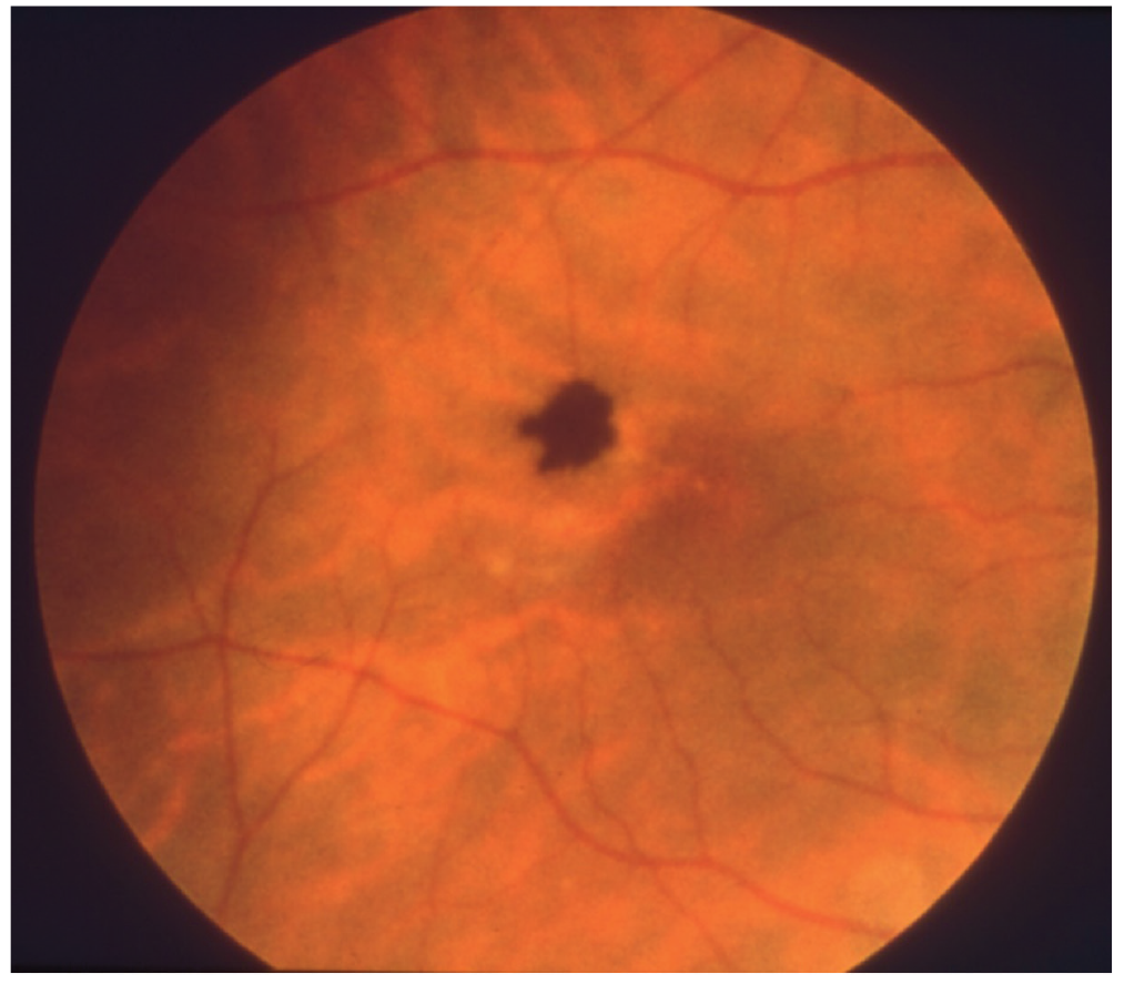
Genellikle unilateral ve koyu siyah renkli bir tümör olarak ortaya çıkar. Düzensiz sınırlıdır. 0,5-1 mm boyutlarında siyah bir nodüldür. Makülayı tutar ().34,35 Hafifçe dilate olmuş besleyici arteriyol ve drene eden venülü mevcuttur. Görme genellikle korunmuştur. Olguların çoğunda lezyon etrafında retinal traksiyon mevcuttur (%80).34,35 Vitreusta pigmente hücreler görülebilir (%20).35 Lezyon ile ilişkili tam kat maküler hol gelişimi bildirilmiştir.36 Görme kaybı fovea traksiyonu ve santral fovea tutulumu neticesinde ortaya çıkar.35
İnceleme Yöntemleri
Ultrasonografik incelemede CSHRPE lezyonları orta iç reflektivitede nodüler lezyon olarak görülür.35 FAF’da hipootofloresans görünüm izlenir.37 OKT’de yüzeyi hiperreflektif görünümde olan, tam kat retina tutulumu ve koroid gölgelenmesi gösteren lezyon izlenir.37,38 OKTA’da tümör içi damarlar tesbit edilebilir.39,40 FA’da erken fazda başlayıp tüm anjiyografi boyunca devam eden hipofloresans görünüm izlenir ve sızıntı görülmez.35,38
Tedavi ve Prognoz
Lezyon ilerleyici olmadığından genelde tedaviye gerek yoktur. Periyodik gözlem önerilir. Makülayı ilgilendiren lezyonlarda görme kaybı kaçınılmazdır. Maküler hol, traksiyon yapan epiretinal membran gelişmesi durumunda vitreoretinal cerrahi uygulanabilir.36
Retina ve Retina Pigment Epitelinin Kombine Hamartomu (CHRRPE)
Klinik Özellikler
CHRRPE nadir görülen iyi huylu bir lezyondur.41,42,43 Görülme yaşı 1-74 arasında olup, ortalama tanı yaşı 23’tür.44 Kombine hamartom genellikle unilateraldir.24 Çoğu olgu sporadiktir.24 Kombine hamartomun, nörofibromatozis Tip 2’li hastalarda artmış oranda görüldüğü bildirilmiştir. Daha nadir hastalıklar arasında nörofibromatozis Tip 1, Poland sendromu, Gorlin sendromu, brankiyookülofasiyal sendrom sayılabilir.24 Kombine hamartom, vasküler, glial ve pigment epitel komponentlerinden oluşur.
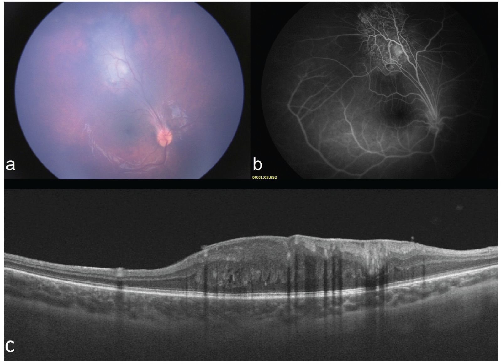
Optik diskte veya fundusun diğer bölgelerinde görülebilir (). CHRRPE yerleşimine göre peripapiller, maküler ve periferik olarak üç grupta incelenir. CHRRPE’nin iç retinadan kaynaklandığı, zamanla dış retinaya doğru ilerlediği ve tümörün lokalizasyonundan bağımsız olarak maküla kalınlığında artış görülebileceği düşünülmektedir.44 Genç hastalarda iç retina ağırlıklı olmak üzere kısmi tutulum, yaşlı hastalarda ise tam kat retina tutulumu daha çok görülür. Preretinal fibrozis genç hastalarda, pigmenter değişiklikler ise yaşlı hastalarda daha çok tesbit edilir. Ortalama maküla kalınlığında artış maküla yerleşimli lezyonlarda diğer lokalizasyondaki lezyonlara göre daha sık görülür.44 Retinanın anatomik durumuna göre CHRRPE’nin üç evresi vardır: 1) retina traksiyonu yok, 2) retina traksiyonu veya retinoskizis mevcut, 3) retina dekolmanı var.45 Peripapiller CHRRPE lezyonlarında pigmentasyon, tam kat retina tutulumu, intraretinal kistik boşluklar, ellipsoid zon/RPE hasarı ve koroid neovaskülarizasyonu maküler CHRRPE lezyonlarına kıyasla daha fazla görülür.46 Görme kaybı, optik disk, papillomaküler demet ve fovea tutulumuna bağlıdır.41,42 Epiretinal membran formasyonuna bağlı makülada traksiyonel distorsiyon gelişir. Genellikle malign transformasyon göstermez. Koroid neovaskülarizasyonu, vitreus hemorajisi, retinoskizis ve maküler hole neden olabilir.47
İnceleme Yöntemleri
Kombine hamartomun tanısı indirekt oftalmoskopi ile konur. Ultrasonografik incelemede CHRRPE lezyonları hafif kabarık, orta-yüksek iç reflektivitede, akustik solid lezyon olarak görülür. Peripapiller pigmente lezyonlar FAF’da hiperotofloresans gösterirler.46 OKT’de lezyonun anatomisine göre CHRRPE üç gruba ayrılır: A) sadece epiretinal komponent; B) kısmı retina tutulumu; C) tam retina ve RPE tutulumu.45 Patogenezde fokal vitreoretinal traksiyon mevcuttur. İç retinada “testere dişi” (mini peak) ve “omega işareti” (maksi peak) görünümü izlenebilir.48,49 “Testere dişi” ve “omega işareti” bulguları genellikle genç hastalarda ortaya çıkar ().44 OKTA’da retina damarlarında değişiklikler ve “filigree” (telkari) paternde tümör içi damarlar görülür.50,51 FA’da hiperfloresans bir lezyondur (). RPE’de artmış pigmentasyon olsa da RPE atrofisi ve hücre göçü nedeniyle erken ve geç hiperfloresans izlenir. Mikroanevrizma ve telenjiektazik damarlar saptanır. Telenjiektazik damarlardan genelde sızıntı izlenmese de geç fazda tortuoz damarlardan minimal sızıntı görülebilir.51,52
Tedavi ve Prognoz
Tedavide periyodik izlem önerilir. Epiretinal membran soyulması cerrahisi tartışmalıdır. Bazı olgularda vitrektomi, membran soyma cerrahisi ile görme artışı bildirilmiştir.53,54 Sekonder koroid neovaskülarizasyonu gelişmişse intravitreal anti-VEGF enjeksiyonu yapılabilir.55 Ambliyopi tedavisi de yapılmalıdır.24 Ancak nadiren görme artışı sağlanabilmektedir. Olguların çoğunda görme kaybı gelişir.24
Retina Hemanjiyoblastomu (Retina Kapiller Hemanjiyomu)
Klinik Özellikler
Retina hemanjiyoblastomu iyi huylu vasküler bir tümördür.56 Tipik olarak periferik retina veya optik diskte yerleşen kırmızı-pembe renkli tümördür (). Kıvrımlı, dilate olmuş besleyici ve drene edici damarları vardır.56 Tek veya multipl olabilir. Hastalığın iki tipi mevcuttur: eksüdatif ve traksiyonel.56 Eksüdatif tipte intraretinal ve subretinal eksüdasyon; traksiyonel tipte retinada gliozis, vitreoretinal traksiyon, vitreus hemorajisi ve traksiyonel retina dekolmanı mevcuttur. Ekzofitik ve endofitik büyüme gösterebilir.56
İzole veya VHL sendromunun bir parçası olarak görülebilir. Soliter retina hemanjiyoblastomu mevcut olan <10 yaş olgularda VHL sendromu gelişme riski %45, >60 yaş olgularda ise <%1’dir.57 VHL sendromunda feokromositoma, renal hücreli karsinom, santral sinir sistemi hemanjiyoblastomları, pankreas kistleri ve nöroendokrin tümörler saptanabilir. VHL Alliance, VHL sendromunda 30 yaşına kadar 6-12 aylık aralıklarla ve 30 yaşından sonra ise yıllık dilate fundus muayenesi önermektedir (https://www.vhl.org/patients/clinical-care/screening). VHL sendromunun ilk bulgusu sıklıkla retina hemanjiyoblastomudur (%50). VHL sendromu olan hastalarda retina hemanjiyoblastomu ortalama 25 yaşında görülür.58 Olguların yaklaşık %58’i bilateraldir.59 Yirmi yaşından büyük hastalarda santral sinir sistemi hemanjioblastomu, >40 yaş hastalarda feokromasitoma, >50 yaş hastalarda renal hücreli karsinom görülür.58 En sık ölüm nedeni renal hücreli karsinomdur.58
İnceleme Yöntemleri
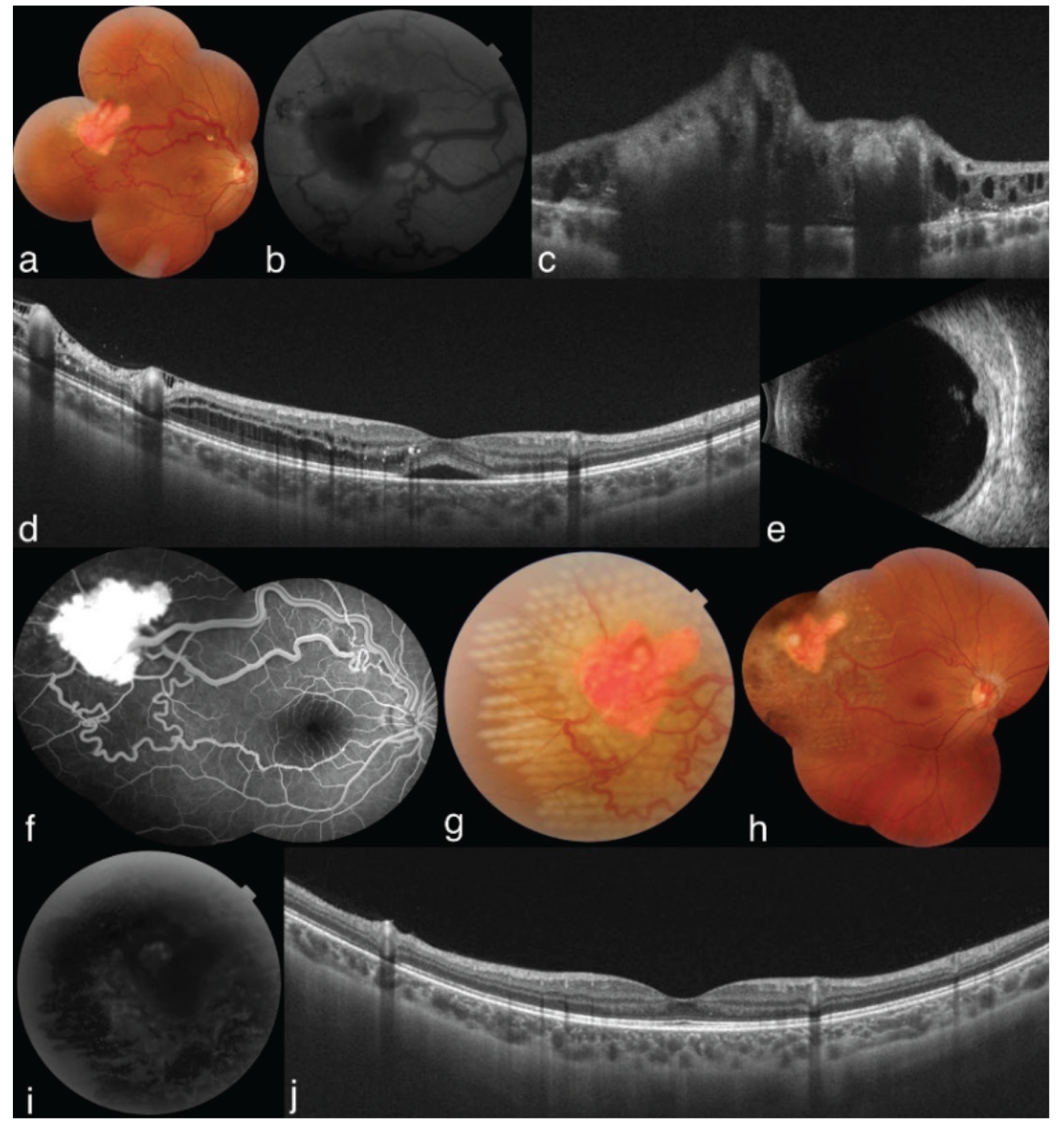
FAF’da hipootofloresans lezyon görülür (). OKT’de retinadan kaynaklanan yüksek reflektif lezyon, dış retinada kompresyon, retina ödemi ve lokalize retina dekolmanı izlenir (). Ultrasonografik incelemede retina hemanjiyoblastomu kabarık, orta-yüksek iç reflektivite gösteren lezyon olarak görülür (). FA’da lezyon arteriyel fazda hiperfloresans gösterir, hiperfloresans geç fazda artar ve vitreus içine boya sızıntısı olur (). Besleyici ve drene edici damarlar OKTA ile FA’ya göre daha iyi tesbit edilebilir.60 Çünkü OKTA’da sızıntı ve göllenme izlenmez. Ama OKTA ile periferik tümörlerin görüntülenmesi mümkün değildir.60,61
Tedavi ve Prognoz
Bazı küçük asemptomatik kitlelerde periyodik izlem yeterli olabilir ve bazen spontan regresyon izlenebilir.62 Sınırlı retina eksüdasyonu veya retina dekolmanı olan tümörlerde laser fotokoagülasyon (), kriyoterapi, transpupiller termoterapi, fotodinamik tedavi, daha ileri tümörlerde ise plak radyoterapi veya eksternal ışın tedavisi yapılabilir.63 Başarılı tedavi sonrasında lezyonda küçülme, besleyici damarlarda incelme ve makülada eksüdatif belirtilerde gerileme meydana gelir (). Vitreus traksiyonu ve retina dekolmanı gelişmişse pars plana vitrektomi de uygulanabilir. Bazı tümörlerde endorezeksiyon da denenebilir.64 Yakın zamanda Amerika Birleşik Devletleri Gıda ve İlaç İdaresi (FDA) VHL ilişkili renal hücreli karsinomları, merkezi sinir sistemi hemanjiyoblastomları veya pankreas nöroendokrin tümörleri olan hastalarda hipoksi ile indüklenebilir faktör inhibitörü olan belzutifan kullanımını onaylamıştır (https://www.fda.gov/drugs/resources-information-approved-drugs/fda-approves-belzutifan-cancers-associated-von-hippel-lindau-disease).
Retina Kavernöz Hemanjiyomu
Klinik Özellikler
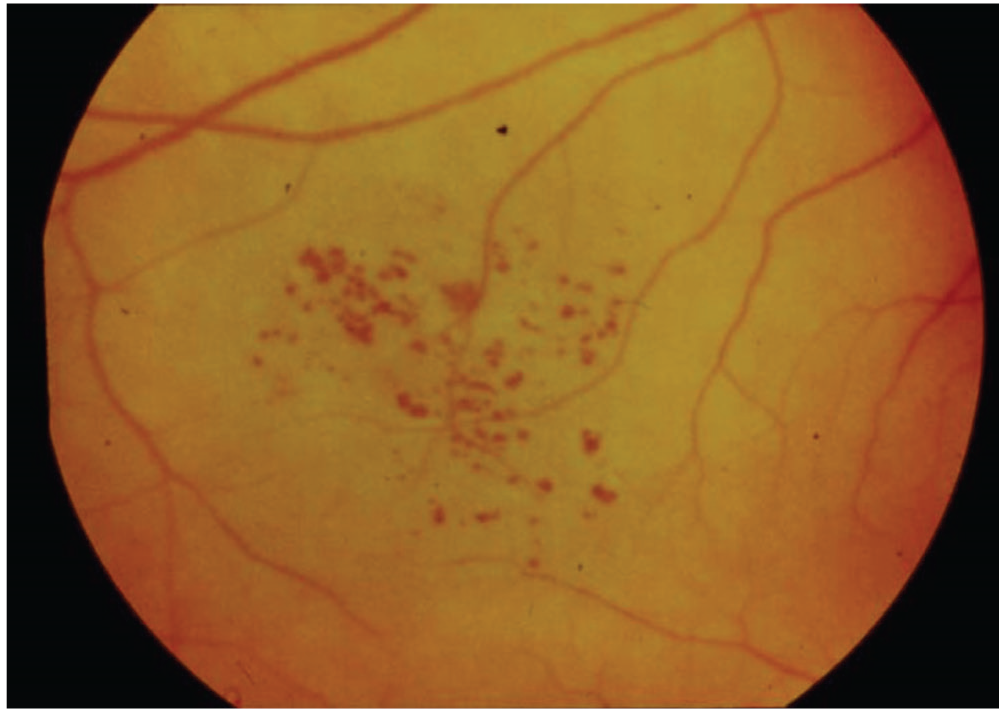
Retina kavernöz hemanjiyomu, unilateral, iyi huylu vasküler hamartomatöz bir lezyondur. Olguların %90’ı unifokaldir.65 Lezyonlar, retinada veya optik sinir başında “üzüm salkımı benzeri” ince duvarlı sakküler anjiyomatöz yapıların oluşumu ile karakterizedir (). Malign transformasyon bildirilmemiştir. Genelde sporadiktir, fakat otozomal dominant şekilde de ortaya çıkabilir. Bu sendromik hastalıkta serebral ve deri hemanjiyomları ile ilişkili olabilir.66 KRIT1/CCM1, CCM2/MGC4607, CCM3/PDCD10 ve 7q mutasyonları görülebilir.67 CCM3 mutasyonu intrakraniyal hemoraji ile ilişkilidir.68
İnceleme Yöntemleri
Ultrasonografik incelemede retina kavernöz hemanjyomu kabarık, yüksek iç reflektivite gösteren lezyon olarak görülür. FA’da erken fazda tümör hipoofloresans görünür ve geç venöz fazda yavaş dolum izlenir. Sakkülün üst yarısında boyanın birikmesi ve altta hipofloresans varlığı “floresein şapkası” görünümü verir.69 Bu patern, eritrositlerin sakkülün dibinde çökmesi ile hipofloresans göstermesine, üstte ise plazma içinde serbest floreseinden kaynaklanan hiperfloresansa bağlıdır.69
Tedavi ve Prognoz
Olguların çoğu tedavi gerektirmez. Tekrarlayan vitreus hemorajisi varsa pars plana vitrektomi uygulanabilir.70 Şiddetli traksiyon ve görme kaybı varsa membran soyulması yapılabilir.71
Sonuçlar
Retina ve optik diskin hamartomları arasında glial hücre kaynaklı astrositik hamartom, RPE ve retinadan kaynaklanan CHRPE, CSHRPE, CHRRPE ve vasküler tümörler olan retina hemanjiyoblastomu ve retina kavernöz hemanjiyomu yer almaktadır. Bu lezyonların çoğu asemptomatiktir ve rutin göz muayenesi için başvuran hastalarda rastlanır. Maküla yerleşimli lezyonlar görme keskinliğinde azalma, görme alanı kaybı gibi bulgulara yol açabilir.
Retina ve optik disk hamartomları izole olabildiği gibi sistemik hastalıklarla ilişkili de olabilir. Sistemik hastalıkların başlangıç bulgusu olabileceğinden bu hamartomların hangi sendrom/hastalıklarla görüldüğünün göz doktoru tarafından bilinmesi gerekmektedir.
Retina ve optik disk hamartomlarının çoğu tedavi gerektirmez. Periyodik gözlem yapılmalıdır. Genelde iyi huylu ve yavaş büyüyen lezyonlardır, nadiren malign transformasyon gelişebilir. Tümöre sekonder komplikasyonlar gelişirse tedavi uygulanabilir.
Dipnotlar
Etik
Hakem Değerlendirmesi
Hakem Değerlendirmesi: Editörler kurulu dışında olan kişiler tarafından değerlendirilmiştir.
Yazarlık Katkıları
Cerrahi ve Medikal Uygulama: I.M., A.K.G., Konsept: I.M., A.K.G., Dizayn: I.M., A.K.G., Veri Toplama veya İşleme: I.M., A.K.G., Analiz veya Yorumlama: I.M., A.K.G., Literatür Arama: I.M., A.K.G., Yazan: I.M., A.K.G.
Çıkar Çatışması
Çıkar Çatışması: Yazarlar tarafından çıkar çatışması bildirilmemiştir.
Finansal Destek: Yazarlar tarafından finansal destek almadıkları bildirilmiştir.
Astrocytic Hamartomas of the Retina and Optic Disc
Clinical Features
Astrocytic hamartomas of the retina and optic disc are benign lesions. They are most commonly seen in patients with TSC. They can also occur in isolation or secondary to other diseases. Approximately 50% of patients with TSC have optic disc and retinal astrocytic hamartoma, and approximately 30% of TSC patients with astrocytic hamartoma develop bilateral tumors. Astrocytic hamartoma is less commonly associated with neurofibromatosis type 1, retinitis pigmentosa, Usher syndrome, and Stargardt disease.
Astrocytic hamartoma develops from glial cells. Tumorgenesis is caused by mutations in the TSC1 and TSC2 genes, which encode hamartin and tuberin, respectively., Retinal astrocytic hamartoma, facial angiofibromas, and depigmented macules on the skin resembling vitiligo are the typical triad of patients with TSC.
Astrocytic hamartoma usually presents as a flat, round, transparent lesion (Figure 1a). The lesion then grows into a nodular structure and becomes calcified., Sometimes the central part of the lesion is calcific, while the peripheral part may be transparent. There may be hard exudates around astrocytic hamartomas. Hard exudates are generally absent in untreated retinoblastoma/retinocytoma., In contrast, changes in the retinal pigment epithelium (RPE) observed in retinoblastomas and retinocytomas do not usually occur in astrocytic hamartomas. Calcification in astrocytic hamartomas is bright yellow with an appearance similar to fish eggs. However, the calcification in retinoblastoma is chalky white. Other diseases that should be considered in the differential diagnosis include optic nerve head drusen, acquired retinal astrocytomas, reactive gliosis, and conditions that cause optic disc edema.
Although astrocytic hamartomas generally have a stable course, giant cell astrocytomas can show progressive growth and cause secondary glaucoma and globe destruction. These tumors are considered malignant but do not metastasize.
Examination Methods
Astrocytic hamartomas appear on ultrasonographic examination as an acoustically solid mass and intralesional calcification is detected. On fundus autofluorescence (FAF) imaging, astrocytic hamartomas demonstrate hyperautofluorescence, depending on their calcium content (Figure 1b). Optical coherence tomography (OCT) shows a dome- or plateau-shaped lesion with moderate to high reflectivity and choroidal shadowing (Figure 1c,d). The choriocapillaris is preserved and “moth-eaten” cavities may be visible. OCT angiography (OCTA) reveals a well-defined, hyperreflective lesion and finely branching tumor vessels., Retinal flow signals within the tumor can be detected on B-mode angiography (Figure 1e). The tumor vasculature is seen in the superficial and deep retina (Figure 1f,g). The outer retina and choriocapillaris show hyporeflective changes due to shadowing/masking caused by calcium or high blood flow within the lesion (Figure 1h,i). Because of the moth-eaten cavities within the lesion, areas of nonperfusion are seen in the deep retinal plexus and projection artifacts can be observed in the outer retina and choriocapillaris. The lesion is hyporeflective in the full macular composite image (Figure 1j). Hyperreflective signals in the lesion center are due to the tumor vasculature in the deep capillary plexus. On fluorescein angiography (FA), early blockage can be seen in the choroidal phase, while the intrinsic tumor vessels begin filling and hyperfluorescence gradually increases during the arterial phase. There may be leakage in the late venous phase. The differential diagnosis of astrocytic hamartoma includes retinoblastoma, retinocytoma, myelinated nerve fibers, and massive retinal gliosis.,,
Based on clinical and OCT findings, four types of retinal astrocytic hamartoma have been identified (Pichi classification). Type 1 appears as a flat retinal lesion (<500 µm) on OCT, with no clinical signs of retinal traction. Type 2 appears as a mildly raised retinal lesion (>500 µm) on OCT, with clinical signs of retinal traction. Type 3 appears as a raised lesion (>500 µm) with calcification in the inner retina on OCT and “mulberry-like” calcification is observed clinically. Type 4 appears as a raised lesion with optically hollow cavities on OCT and the clinical appearance is a flat, non-calcified lesion in the inner retina. Type 2 retinal astrocytic hamartoma is significantly more common in TSC patients with fibrous skin plaques, Type 3 in those with subependymal giant cell astrocytic hamartoma, and Type 4 in those with lung lymphangiomyomatosis.
Treatment and Prognosis
Astrocytic hamartoma is generally stable and does not require treatment. Rarely, spontaneous regression may occur. Periodic monitoring is important because of the risk of vitreous hemorrhage, retinal exudation, retinal detachment, and neovascular glaucoma. Laser photocoagulation can be applied to small lesions. Photodynamic therapy can be attempted for tumors that are larger and symptomatic (if exudates and fluid are present). Anti-vascular endothelial growth factor (anti-VEGF) injections can be administered in cases with secondary choroidal neovascularization. If vitreous hemorrhage from intratumoral fine vessels or neovascularization occurs, pars plana vitrectomy surgery may be required. Giant cell astrocytic hamartoma may show an aggressive course, and enucleation may be necessary due to tumor necrosis, vitreous hemorrhage, subretinal hemorrhage, massive exudation, and the development of neovascular glaucoma. Neovascular glaucoma occurs as a result of intratumoral necrosis or chronic retinal detachment. TSC patients with subependymal giant cell astrocytoma and renal angiomyolipoma can be treated with m-TOR inhibitors (rapamycin, everolimus, sirolimus).
Congenital Hypertrophy of the Retinal Pigment Epithelium (CHRPE)
These are flat, round, and pigmented fundus lesions that are not raised from the surface. They are located at the RPE level. There are three variants: solitary, multifocal, and atypical (Figure 2a). Histopathologically, CHRPE lesions are composed of hypertrophic RPE cells containing excessive pigment granules. The choriocapillaris and choroid are normal. The photoreceptor layer over the RPE may be normal or atrophic. Photoreceptor atrophy fully manifests as the lesion becomes chronic. Patients with photoreceptor atrophy exhibit absolute scotomas on visual field examination.
The margins of CHRPE lesions are usually smooth but can occasionally be irregular. They often occur in the peripheral fundus; peripapillary localization is less common. There may be depigmented lacunae within the lesion and a hypopigmented halo around the lesion (Figure 2a). CHRPE lesions can show minimal growth, especially in myopic eyes. Rarely, lesions such as RPE adenoma/adenocarcinoma may develop from CHRPE.
CHRPEs are occasionally amelanotic. These types of lesions are called amelanotic CHRPE. The retina and retinal vessels overlying the CHRPE are normal. Focal intraretinal pigmentation may be present. In rare cases, neovascularization may occur at the edge of CHRPE.
One type of CHRPE is associated with FAP. The gene responsible for FAP is called the APC (adenomatous polyposis coli) gene, located in the 5q21-q22 region. CHRPEs associated with FAP are generally oval-shaped and have tail-like extensions. These lesions are called pigmented ocular fundus lesions (POFL) to distinguish them from other CHRPEs. Their tails may be depigmented and they may contain lacunae. Histopathologically similar to CHRPE, they have pathological appearances of RPE hypertrophy, hyperplasia, and hamartoma. POFLs usually have a base diameter smaller than 5 mm.
POFL can be associated with Gardner syndrome and Turcot syndrome. In Gardner syndrome, hundreds of polyps appear in the colon and give rise to adenocarcinoma of the colon. Prophylactic colectomy is recommended. Extracolonic cancers in Gardner syndrome occur in the thyroid, adrenal gland, and liver. Benign lesions include head and orbit osteomas, sebaceous cysts, lipomas, and fibromas. Opaque jaw lesions can also be observed. In general, detection of 4 or more POFLs is a strong indicator of FAP. In Turcot syndrome, patients develop brain tumors in addition to POFL.
On ultrasonographic examination, CHRPE lesions appear as flat or minimally raised (<0.5 mm), acoustically solid lesions. FAF imaging typically shows hypoautofluorescence due to the lesions’ high melanin content (Figure 2b). Non-pigmented halos or cavities may show autofluorescence. On OCT they are flat lesions at the RPE level that contain hyperreflective deposits and cause choroidal shadowing (Figure 2c,d). The inner retinal layers are normal, but the outer retinal layers exhibit thinning. On OCTA, CHRPEs are generally well-defined and hyperreflective in the superficial and deep retinal plexi. B-mode angiography demonstrates flow signals in the retina overlying the tumor. Masking of the outer retina and choriocapillaris causes the appearance of “signal void” areas. On FA, leakage is not observed. The underlying choroidal fluorescence is blocked in areas other than depigmented halos or cavities.
CHRPE lesions generally do not require treatment. However, long-term follow-up is essential. Rarely, RPE adenoma/adenocarcinoma may arise from CHRPE. This type of RPE tumor can be confused with choroidal malignant melanoma. RPE tumors are darker brown/near black in color. However, as they are indistinguishable from choroidal malignant melanoma, RPE adenoma/adenocarcinoma is usually treated by enucleation. The use of proton beam therapy for CHRPE-derived RPE adenocarcinoma was reported in one case.
Congenital Simple Hamartoma of the Retinal Pigment Epithelium (CSHRPE)
CSHRPE usually appears as a unilateral and dark black tumor with irregular margins. They are black nodules 0.5-1 mm in size and involve the macula (Figure 3)., A slightly dilated afferent arteriole and efferent venule are present. Vision is generally preserved. There is retinal traction around the lesion in most cases (80%)., Pigmented cells may be detected in the vitreous (20%). The development of full-thickness macular hole associated with CSHRPE has been reported. Loss of vision occurs as a result of foveal traction and central foveal involvement.
On ultrasonographic examination, CSHRPE appears as a nodular lesion with moderate internal reflectivity. The lesion is hypoautofluorescent on FAF. OCT demonstrates a lesion with a hyperreflective surface that shows full-thickness retinal involvement and choroidal shadowing., Intratumoral vessels can be detected on OCTA., On FA, there is hypofluorescence starting in the early phase and continuing throughout the entire angiography, with no leakage.,
As the lesion is not progressive, treatment is generally not necessary but periodic monitoring is recommended. Vision loss is inevitable with lesions involving the macula. Vitreoretinal surgery can be performed if macular hole or tractional epiretinal membrane develop.
Combined Hamartoma of the Retina and Retinal Pigment Epithelium (CHRRPE)
CHRRPE is a rare benign lesion.,,, It presents between the ages of 1 and 74 years and the mean age at diagnosis is 23 years. Combined hamartoma is usually unilateral and most cases are sporadic. It has been reported to occur more frequently in patients with neurofibromatosis type 2. Less common associations include neurofibromatosis type 1, Poland syndrome, Gorlin syndrome, and branchio-oculo-facial syndrome. Combined hamartoma consists of vascular, glial, and pigment epithelial components.
It can occur in the optic disc or other areas of the fundus (Figure 4a). CHRRPE is classified into three groups according to location: peripapillary, macular, and peripheral. CHRRPE is believed to originate from the inner retina and progress towards the outer retina over time, and an increase in macular thickness may occur regardless of tumor location. Young patients exhibit partial involvement, mainly of the inner retina, and full-thickness retinal involvement is more often seen in older patients. Preretinal fibrosis is more common in young patients, while pigmentary changes are more frequently detected in older patients. An increase in average macular thickness is more common in macular lesions than with those in other locations. There are three stages of CHRRPE according to the anatomical condition of the retina: 1) no retinal traction, 2) retinal traction or retinoschisis is present, and 3) retinal detachment is present. Pigmentation, full-thickness retinal involvement, intraretinal cystic cavities, ellipsoid zone/RPE disruption, and choroidal neovascularization are more common in peripapillary CHRRPE lesions compared to macular CHRRPE lesions. Vision loss varies depending on optic disc, papillomacular bundle, and foveal involvement., Tractional distortion occurs in the macula due to epiretinal membrane formation. They usually do not show malignant transformation. Choroidal neovascularization may cause vitreous hemorrhage, retinoschisis, and macular hole.
Combined hamartoma is diagnosed by indirect ophthalmoscopy. On ultrasonographic examination, CHRRPE lesions appear as slightly raised, acoustically solid lesions with moderate to high internal reflectivity. Peripapillary pigmented lesions exhibit hyperautofluorescence on FAF. On OCT, CHRRPE is divided into three groups according to lesion anatomy: A) epiretinal component only, B) partial retinal involvement, and C) full retinal and RPE involvement. The pathogenesis involves focal vitreoretinal traction. The inner retina exhibits a “sawtooth” pattern (mini-peak) and “omega sign” (maxi-peak)., The sawtooth and omega signs are usually detected in young patients (Figure 4c). OCTA demonstrates retinal vascular alterations and a “filigree” pattern in the intratumoral vessels., On FA, the lesion is hyperfluorescent (Figure 4b). Despite increased pigmentation in the RPE, early and late hyperfluorescence is observed because of RPE atrophy and cell migration. Microaneurysms and telangiectatic vessels are observed. Although there is usually no leakage from the telangiectatic vessels, minimal leakage from the tortuous vessels may be seen in the late phase.,
Periodic monitoring is recommended for treatment. The role of epiretinal membrane peeling surgery is controversial. In some cases, visual improvement has been reported with vitrectomy and membrane peeling., Intravitreal anti-VEGF injection can be administered in eyes with secondary choroidal neovascularization. Amblyopia treatment should also be provided. However, visual improvement is rarely achieved. Vision loss occurs in most cases.
Retinal Hemangioblastoma (Retinal Capillary Hemangioma)
Retinal hemangioblastomas are benign vascular tumors. They are red-pink tumors typically located in the peripheral retina or optic disc (Figure 5a). Their afferent and efferent vessels are tortuous and dilated. They may be single or multiple. The disease has two types, exudative and tractional. The exudative type is characterized by intraretinal and subretinal exudation, the tractional type by retinal gliosis, vitreoretinal traction, vitreous hemorrhage, and tractional retinal detachment. They may exhibit exophytic and endophytic growth.
Retinal hemangioblastomas may be isolated or occur as part of VHL syndrome. Patients presenting with solitary retinal hemangioblastoma before the age of 10 years have a 45% risk of developing VHL syndrome, while the risk is <1% in those over 60 years of age. Pheochromocytoma, renal cell carcinoma, central nervous system hemangioblastomas, pancreatic cysts, and neuroendocrine tumors may be seen in VHL syndrome. The VHL Alliance recommends dilated fundus examination every 6-12 months until the age of 30 and annually after the age of 30 for patients with VHL syndrome (https://www.vhl.org/patients/clinical-care/screening). Retinal hemangioblastoma is often the initial sign of VHL syndrome (50%) and is seen in VHL patients at an average age of 25 years. Approximately 58% of cases are bilateral. Central nervous system hemangioblastoma is seen in patients aged >20 years, pheochromocytoma in patients aged >40 years, and renal cell carcinoma in patients aged >50 years. Renal cell carcinoma is the most common cause of death.
FAF shows a hypoautofluorescent lesion (Figure 5b). On OCT, a hyperreflective lesion originating from the retina with compression of the outer retina, retinal edema, and localized retinal detachment is observed (Figure 5c,d). On ultrasonographic examination, retinal hemangioblastoma appears as a raised lesion with moderate to high internal reflectivity (Figure 5e). On FA, the lesion shows hyperfluorescence in the arterial phase that increases in the late phase, with dye leakage into the vitreous (Figure 5f). The afferent and efferent vessels are better detected with OCTA than with FA, because leakage and pooling are not seen on OCTA. However, peripheral tumors cannot be imaged with OCTA.,
For some small, asymptomatic masses, periodic monitoring can be sufficient and spontaneous regression may be observed in some cases. Tumors with limited retinal exudation or retinal detachment can be treated with laser photocoagulation (Figure 5g,i), cryotherapy, transpupillary thermotherapy, and photodynamic therapy and advanced tumors may be treated with plaque radiotherapy or external beam therapy. Successful treatment results in shrinkage of the lesion, narrowing of the afferent vessels, and regression of exudative symptoms in the macula (Figure 5h,j). If vitreous traction and retinal detachment occur, pars plana vitrectomy can also be performed. Endoresection may also be attempted for some tumors. The United States Food and Drug Administration (FDA) recently approved the use of belzutifan, a hypoxia-inducible factor inhibitor, in patients with VHL-associated renal cell carcinomas, central nervous system hemangioblastomas, or pancreatic neuroendocrine tumors (https://www.fda.gov/drugs/resources-information-approved-drugs/fda-approves-belzutifan-cancers-associated-von-hippel-lindau-disease).
Retinal Cavernous Hemangioma
Retinal cavernous hemangioma is a unilateral, benign vascular hamartomatous lesion. Ninety percent of cases are unifocal. The lesions are characterized by the formation of thin-walled saccular angiomatous structures in the retina or optic nerve head that resemble a bunch of concord grapes (Figure 6). Malignant transformation has not been reported. It is usually sporadic but can also show autosomal dominant inheritance. In this syndromic association, it may co-exist with cerebral and skin hemangiomas. KRIT1/CCM1, CCM2/MGC4607, CCM3/PDCD10, and 7q mutations may be present. The CCM3 mutation is associated with intracranial hemorrhage.
On ultrasonographic examination, retinal cavernous hemangioma appears as a raised lesion with high internal reflectivity. On FA, the tumor exhibits hypofluorescence in the early phase and slow filling in the late venous phase. Dye accumulation in the upper half of the saccule and the presence of hypofluorescence underneath give the appearance of a “fluorescein cap.” This pattern is the result of hypofluorescence caused by erythrocyte sedimentation at the bottom of the saccule and hyperfluorescence caused by free fluorescein in the plasma at the top.
Most cases do not require treatment. Recurrent vitreous hemorrhage can be treated with pars plana vitrectomy. Membrane peeling can be performed in cases with severe traction and vision loss.
Kaynakça
Batsakis JG. Pathology consultation. Nomenclature of developmental tumors. Ann Otol Rhinol Laryngol. 1984;93:98-99.
Ali SA, Mulita F. Hamartoma. In: StatPearls [Internet]. Treasure Island (FL): StatPearls Publishing; 2022.
Lu DS, Karas PJ, Krueger DA, Weiner HL. Central nervous system manifestations of tuberous sclerosis complex. Am J Med Genet C Semin Med Genet. 2018;178:291-298.
Karaconji T, Whist E, Jamieson RV, Flaherty MP, Grigg JRB. Neurofibromatosis Type 1: Review and Update on Emerging Therapies. Asia Pac J Ophthalmol (Phila). 2019;8:62-72.
Asthagiri AR, Parry DM, Butman JA, Kim HJ, Tsilou ET, Zhuang Z, Lonser RR. Neurofibromatosis type 2. Lancet. 2009;373:1974-1986.
Wiley HE, Krivosic V, Gaudric A, Gorin MB, Shields C, Shields J, Aronow ME, Chew EY. Management Of Retınal Hemangıoblastoma In Von Hıppel-Lındau Dısease. Retina. 2019;39:2254-2263.
Shields JA, Shields CL. Tumors and related lesions of the pigmented epithelium. Asia Pac J Ophthalmol (Phila). 2017;6:215-223.
Hodgson N, Kinori M, Goldbaum MH, Robbins SL. Ophthalmic manifestations of tuberous sclerosis: a review. Clin Exp Ophthalmol. 2017;45:81-86.
Johnson MW, Kerfoot C, Bushnell T, Vinters HV. Hamartin and tuberin expression in human tissues. Mod Pathol. 2001;14:202-210.
van Slegtenhorst M, Nellist M, Nagelkerken B, Cheadle J, Snell R, van den Ouweland A, Reuser A, Sampson J, Halley D, van der Sluijs P. Interaction between hamartin and tuberin, the TSC1 and TSC2 gene products. Hum Mol Genet. 1998;7:1053-1057.
Zimmer-Galler IE, Robertson DM. Long-term observation of retinal lesions in tuberous sclerosis. Am J Ophthalmol. 1995;119:318-324.
Nyboer JH, Robertson DM, Gomez MR. Retinal lesions in tuberous sclerosis. Arch Ophthalmol. 1976;94:1277-1280.
Giles J, Singh AD, Rundle PA, Noe KP, Rennie IG. Retinal astrocytic hamartoma with exudation. Eye (Lond) 2005;19:724-725.
Shields CL, Manalac J, Das C, Saktanasate J, Shields JA. Review of spectral domain-enhanced depth imaging optical coherence tomography of tumors of the retina and retinal pigment epithelium in children and adults. Indian J Ophthalmol. 2015;63:128-132.
Shields JA, Shields CL, Shields JA, Shields CL, eds. Glial tumors of the retina and optic disc. Intraocular Tumors: an Atlas and Textbook (3rd ed). Lippincott Williams & Wilkins; Philadelphia, PA, USA. 2016:1137–1187.
Gündüz AK, Mirzayev I, Kasimoglu R, Özalp Ateş FS. Swept-source optical coherence tomography angiography findings in choroidal and retinal tumors. Eye (Lond). 2021;35:4-16.
Toledo JJ, Asencio M, García JR, Morales LA, Tomkinson C, Cajigal C. OCT Angiography: Imaging of Choroidal and Retinal Tumors. Ophthalmol Retina. 2018;2:613-622.
Mirzayev I, Gündüz AK, Biçer Ö, Tarlan B. The Final Diagnosis: Retinoblastoma or Pseudoretinoblastoma. J Pediatr Ophthalmol Strabismus. 2021;58:161-167.
Pichi F, Massaro D, Serafino M, Carrai P, Giuliari GP, Shields CL, Veronese C, Ciardella AP, Nucci P. Retınal Astrocytıc Hamartoma: Optical Coherence Tomography Classification and Correlation With Tuberous Sclerosis Complex. Retina. 2016;36:1199-1208.
Şekeroğlu MA, Kıratlı H. Spontaneous regression of presumed peripapillary retinal astrocytic hamartoma. J Fr Ophtalmol. 2021;44:e255-e257.
Gündüz K, Eagle RC Jr, Shields CL, Shields JA, Augsburger JJ. Invasive giant cell astrocytoma of the retina in a patient with tuberous sclerosis. Ophthalmology. 1999;106:639-642. Erratum in: Ophthalmology. 2000;107:413.
MacKeigan JP, Krueger DA. Differentiating the mTOR inhibitors everolimus and sirolimus in the treatment of tuberous sclerosis complex. Neuro Oncol. 2015;17:1550-1559.
Shields CL, Mashayekhi A, Ho T, Cater J, Shields JA. Solitary congenital hypertrophy of the retinal pigment epithelium: clinical features and frequency of enlargement in 330 patients. Ophthalmology. 2003;110:1968-1976.
Shields JA, Shields CL, Shields JA, Shields CL, eds.Tumors and related lesions of the pigment epithelium. Intraocular Tumors: an Atlas and Textbook (3rd ed). Lippincott Williams & Wilkins; Philadelphia, PA, USA. 2016: 1188-1319.
Shields JA, Shields CL, Singh AD. Acquired tumors arising from congenital hypertrophy of the retinal pigment epithelium. Arch Ophthalmol. 2000;118:637-641.
Shields CL, Mashayekhi A, Ho T, Cater J, Shields JA. Solitary congenital hypertrophy of the retinal pigment epithelium: clinical features and frequency of enlargement in 330 patients. Ophthalmology. 2003;110:1968-1976.
Bellamy JP, Cohen SY. Congenital hypertrophy of the retinal pigment epithelium complicated by choroidal neovascularization. Ophthalmol Retina. 2022;6:511.
Shields CL, Pirondini C, Bianciotto C, Harmon SA, Shields JA. Autofluorescence of congenital hypertrophy of the retinal pigment epithelium. Retina. 2007;27:1097-1100.
Shields JA, Shields CL, Shah PG, Pastore DJ, Imperiale SM Jr. Lack of association among typical congenital hypertrophy of the retinal pigment epithelium, adenomatous polyposis, and Gardner’s syndrome. Ophthalmology. 1992;99:1709-1713.
Heinemann MH, Baker RH, Miller HH, DeCosse JJ. Familial polyposis coli: the spectrum of ocular and other extracolonic manifestations. Graefes Arch Clin Exp Ophthalmol. 1991;229:213-218.
Munden PM, Sobol WM, Weingeist TA. Ocular findings in Turcot syndrome (glioma-polyposis). Ophthalmology 1991;98:111-114.
Traboulsi EI, Apostolides J, Giardiello FM, Krush AJ, Booker SV, Hamilton SR, Hussels IE. Pigmented ocular fundus lesions and APC mutations in familial adenomatous polyposis. Ophthalmic Genet. 1996;17:167-174.
Moulin AP, Zografos L, Schalenbourg A. RPE adenocarcinoma arising from a congenital hypertrophy of the RPE (CHRPE) treated with proton therapy. Klin Monatsbl Augenheilkd. 2014;231:411-413.
Laqua H. Tumors and tumor-like lesions of the retinal pigment epithelium. Ophthalmologica. 1981;183:34-38.
Shields CL, Shields JA, Marr BP, Sperber DE, Gass JD. Congenital simple hamartoma of the retinal pigment epithelium: a study of five cases. Ophthalmology. 2003;110:1005-1011.
Stavrakas P, Vachtsevanos A, Karakosta E, Kozeis N, Triantafylla M, Tranos P. Full-thickness macular hole associated with congenital simple hamartoma of retinal pigment epithelium (CSHRPE). Int Ophthalmol. 2018;38:2179-2182.
Nakatsuka AS, Banaee T, Loucks E, El-Annan J. Simple congenital hamartoma of the retinal pigment epithelium. J Ophthalmic Vis Res. 2020;15:261-263.
Teke MY, Ozdal PÇ, Batioglu F, Elgin U, Oztürk F. Congenital simple hamartoma of retinal pigment epithelium: clinical and imaging findings. Case Rep Ophthalmol Med. 2012;2012:654502.
Arjmand P, Elimimian EB, Say EAT, Shields CL. Optical coherence tomography angiography of congenital simple hamartoma of the retinal pigment epithelium. Retin Cases Brief Rep. 2019;13:357-360.
Zola M, Ambresin A, Zografos L. Optıcal Coherence Tomography Angıography Imagıng Of A Congenıtal Sımple Hamartoma Of The Retınal Pıgment Epıthelıum. Retin Cases Brief Rep. 2021;15:289-293.
Gass JD. An unusual hamartoma of the pigment epithelium and retina simulating choroidal melanoma and retinoblastoma. Trans Am Ophthalmol Soc. 1973;71:171-183; discussion 184-185.
Schachat AP, Shields JA, Fine SL, Sanborn GE, Weingeist TA, Valenzuela RE, Brucker AJ. Combined hamartomas of the retina and retinal pigment epithelium. Ophthalmology. 1984;91:1609-1615.
Ticho BH, Egel RT, Jampol LM. Acquired combined hamartoma of the retina and pigment epithelium following parainfectious meningoencephalitis with optic neuritis. J Pediatr Ophthalmol Strabismus. 1998;35:116-118.
Ledesma-Gil G, Essilfie J, Gupta R, Fung AT, Lupidi M, Pappuru RR, Nayak S, Sahoo NK, Kaliki S, Yannuzzi LA, Reid K, Lim L, Sacconi R, Dave V, Singh SR, Ayachit A, Gabrielle PH, Cai S, Lima LH, Querques G, Arevalo JF, Freund KB, Shields CL, Chhablani J. Presumed Natural History of Combined Hamartoma of the Retina and Retinal Pigment Epithelium. Ophthalmol Retina. 2021;5:1156-1163.
Dedania VS, Ozgonul C, Zacks DN, Besirli CG. Novel Classıfıcatıon System For Combıned Hamartoma Of The Retına And Retınal Pıgment Epıthelıum. Retina. 2018;38:12-19.
Gupta R, Fung AT, Lupidi M, Pappuru RR, Nayak S, Sahoo NK, Kaliki S, Yannuzzi L, Reid K, Lim L, Sacconi R, Dave V, Singh SR, Ayachit A, Gabrielle PH, Cai S, Lima LH, Querques G, Arevalo JF, Freund KB, Shields CL, Chhablani J. Peripapillary Versus Macular Combined Hamartoma of the Retina and Retinal Pigment Epithelium: Imaging Characteristics. Am J Ophthalmol. 2019;200:263-269.
Schathat AP, Glaser BM. Retinal hamartoma, acquired retinoschisis, and retinal hole. Am J Ophthalmol. 1985;99:604-605.
Arepalli S, Pellegrini M, Ferenczy SR, Shields CL. Combined hamartoma of the retina and retinal pigment epithelium: findings on enhanced depth imaging optical coherence tomography in eight eyes. Retina. 2014;34:2202-2207.
Chawla R, Kumar V, Tripathy K, Kumar A, Venkatesh P, Shaikh F, Vohra R, Molla K, Verma S. Combined Hamartoma of the Retina and Retinal Pigment Epithelium: An Optical Coherence Tomography-Based Reappraisal. Am J Ophthalmol. 2017;181:88-96.
Gupta R, Pappuru RR, Fung KAT, Lupidi M, Kaliki S, Yannuzzi L, Freund KB, Reid K, Zur D, Iglicki M, Chhablani J. Filigree Vascular Pattern in Combined Hamartoma of Retina and Retinal Pigment Epithelium on OCT Angiography. Ophthalmol Retina. 2019;3:879-887.
Scupola A, Grimaldi G, Sammarco MG, Sasso P, Marullo M, Blasi MA. Multimodal imaging evaluation of combined hamartoma of the retina and retinal pigment epithelium. Eur J Ophthalmol. 2020;30:595-599.
Gündüz AK, Mirzayev I, Tetik D. Wide-field Fundus Imaging and Fluorescein Angiography Findings in Various Pseudoretinoblastoma Conditions. J Pediatr Ophthalmol Strabismus. 2022:1-15.
Stallman JB. Visual improvement after pars plana vitrectomy and membrane peeling for vitreoretinal traction associated with combined hamartoma of the retina and retinal pigment epithelium. Retina. 2002;22:101-104.
Gupta R, Ayachit A, Joshi S, Nayak S, Sahoo NK, Sacconi R, Singh SR, Muftuoglu IK, Querques G, Chhablani J. Optical coherence tomography analysis of surgical outcomes of combined hamartoma of retina and retinal pigment epithelium. Saudi J Ophthalmol. 2021;34:237-242.
Lazzarini TA, Al-Khersan H, Patel NA, Russell JF, Fan KC, De Oliveira G, Negron CI, Mavrofrides E, Berrocal AM. Peripheral combined hamartoma of the retina and retinal pigment epithelium with remote peripapillary choroidal neovascular membrane. Am J Ophthalmol Case Rep. 2020;20:100954.
Shields JA, Shields CL, Shields JA, Shields CL, eds. Vascular tumors of the retina and optic disc. Intraocular Tumors: an Atlas and Textbook, (3rd ed). Lippincott Williams & Wilkins; Philadelphia, PA, USA 2016:1048-1136.
Singh A, Shields J, Shields C. Solitary retinal capillary hemangioma: hereditary (von Hippel-Lindau disease) or nonhereditary? Arch Ophthalmol. 2001;119:232-234. Erratum in: Arch Ophthalmol. 2001;119:1226.
Varshney N, Kebede AA, Owusu-Dapaah H, Lather J, Kaushik M, Bhullar JS. A review of Von Hippel-Lindau syndrome. J Kidney Cancer VHL. 2017;4:20-29.
Wong WT, Agrón E, Coleman HR, Tran T, Reed GF, Csaky K, Chew EY. Clinical characterization of retinal capillary hemangioblastomas in a large population of patients with von Hippel-Lindau disease. Ophthalmology. 2008;115:181-188.
Sagar P, Rajesh R, Shanmugam M, Konana VK, Mishra D. Comparison of optical coherence tomography angiography and fundus fluorescein angiography features of retinal capillary hemangioblastoma. Indian J Ophthalmol. 2018;66:872-876.
Russell JF, Villegas VM, Schwartz SG, Weng CY, Davis JL, Flynn HW Jr, Harbour JW. Multimodal Imaging in the Diagnosis of Exophytic Juxtapapillary Retinal Capillary Hemangioblastoma. Am J Ophthalmol. 2021;225:128-136.
Milewski SA. Spontaneous regression of a capillary hemangioma of the optic disc. Arch Ophthalmol. 2002;120:1100-1101.
Yaman A, Saatci AO, Arikan G, Gunduz K. Involution of endophytic optic disc hemangioma with a single session of photodynamic treatment. Ann Ophthalmol (Skokie). 2007;39:63-66.
Kwan AS, Ramkissoon YD, Gregor ZJ. Surgical management of retinal capillary hemangioblastoma associated with retinal detachment. Retina. 2008;28:1159-1162.
Wang W, Chen L. Cavernous Hemangıoma Of The Retına: A Comprehensive Review of the Literature (1934-2015). Retina. 2017;37:611-621.
Gass JD. Cavernous hemangioma of the retina. A neuro-oculo-cutaneous syndrome. Am J Ophthalmol. 1971;71:799-814.
Reddy S, Gorin MB, McCannel TA, Tsui I, Straatsma BR. Novel KRIT1/CCM1 mutation in a patient with retinal cavernous hemangioma and cerebral cavernous malformation. Graefes Arch Clin Exp Ophthalmol. 2010;248:1359-1361.
Riant F, Bergametti F, Fournier HD, Chapon F, Michalak-Provost S, Cecillon M, Lejeune P, Hosseini H, Choe C, Orth M, Bernreuther C, Boulday G, Denier C, Labauge P, Tournier-Lasserve E. CCM3 Mutations Are Associated with Early-Onset Cerebral Hemorrhage and Multiple Meningiomas. Mol Syndromol. 2013;4:165-172.
Kumar M, Reddy N, Konana VK, Kanakamedla A, Ruia S, Gudimetla J. Fluorescein cap: Fluorescein angiographic feature of retinal cavernous hemangioma. Indian J Ophthalmol. 2018;66:1473-1474.
Haller JA, Knox DL. Vitrectomy for persistent vitreous hemorrhage from a cavernous hemangioma of the optic disk. Am J Ophthalmol. 1993;116:106-107.
Shukla D, Sharan A. Epimacular membrane secondary to an optic nerve head lesion. Br J Ophthalmol. 2012;96:1038-1039, 1048.

